

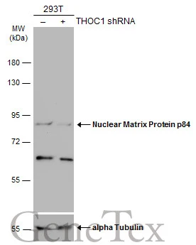
Non-transfected (–) and transfected (+) 293T whole cell extracts (30 μg) were separated by 7.5% SDS-PAGE, and the membrane was blotted with Nuclear Matrix Protein p84 antibody [C1C3] (GTX102919) diluted at 1:2000. The HRP-conjugated anti-rabbit IgG antibody (GTX213110-01) was used to detect the primary antibody.
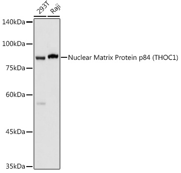
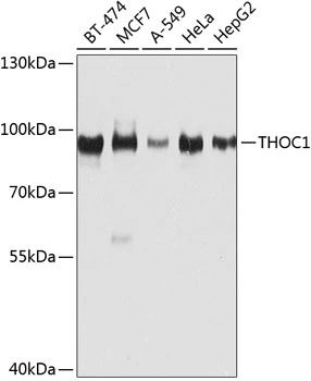
![Various whole cell extracts (30 μg) were separated by 7.5% SDS-PAGE, and the membrane was blotted with Nuclear Matrix Protein p84 antibody [C1C3] (GTX102919) diluted at 1:1000. The HRP-conjugated anti-rabbit IgG antibody (GTX213110-01) was used to detect the primary antibody.](https://www.genetex.com/upload/website/prouct_img/normal/GTX102919/GTX102919_40163_20170825_WB_w_23060119_631.webp "Various whole cell extracts (30 μg) were separated by 7.5% SDS-PAGE, and the membrane was blotted with Nuclear Matrix Protein p84 antibody [C1C3] (GTX102919) diluted at 1:1000. The HRP-conjugated anti-rabbit IgG antibody (GTX213110-01) was used to detect the primary antibody.")

![Nuclear Matrix Protein p84 antibody [C1C3] immunoprecipitates p84 protein in IP experiments. IP Sample: HepG2 whole cell lysate/extract A : 30 μg whole cell lysate/extract of p84 protein expressing HepG2 cells B : Control with 3 μg of pre-immune rabbit IgG C : Immunoprecipitation of p84 by 3 μg of Nuclear Matrix Protein p84 antibody [C1C3] (GTX102919) 7.5% SDS-PAGE The immunoprecipitated p84 protein was detected by Nuclear Matrix Protein p84 antibody [C1C3] (GTX102919) diluted at 1 : 1000. EasyBlot anti-rabbit IgG (HRP) (GTX221666-01) was used as a secondary reagent.](https://www.genetex.com/upload/website/prouct_img/normal/GTX102919/GTX102919_40163_IP_w_23060119_492.webp "Nuclear Matrix Protein p84 antibody [C1C3] immunoprecipitates p84 protein in IP experiments. IP Sample: HepG2 whole cell lysate/extract A : 30 μg whole cell lysate/extract of p84 protein expressing HepG2 cells B : Control with 3 μg of pre-immune rabbit IgG C : Immunoprecipitation of p84 by 3 μg of Nuclear Matrix Protein p84 antibody [C1C3] (GTX102919) 7.5% SDS-PAGE The immunoprecipitated p84 protein was detected by Nuclear Matrix Protein p84 antibody [C1C3] (GTX102919) diluted at 1 : 1000. EasyBlot anti-rabbit IgG (HRP) (GTX221666-01) was used as a secondary reagent.")
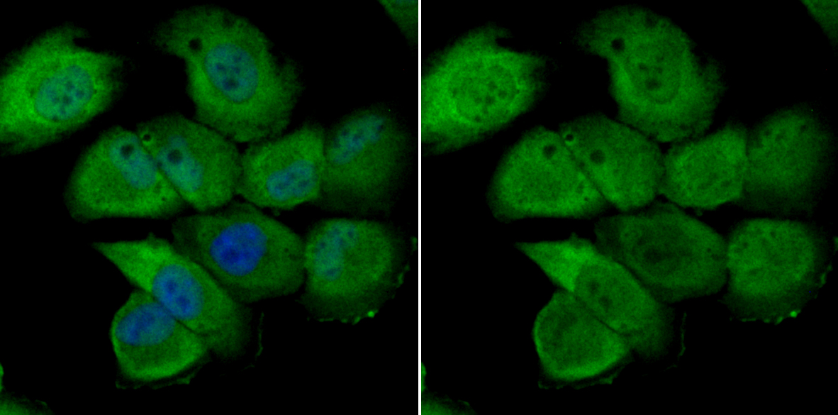
![Nuclear Matrix Protein p84 antibody [C1C3] detects Nuclear Matrix Protein p84 protein at cytoplasm and nucleus by immunofluorescent analysis. Sample: HeLa cells were fixed in 4% paraformaldehyde at RT for 15 min. Green: Nuclear Matrix Protein p84 protein stained by Nuclear Matrix Protein p84 antibody [C1C3] (GTX102919) diluted at 1:500. Blue: Hoechst 33342 staining.](https://www.genetex.com/upload/website/prouct_img/normal/GTX102919/GTX102919_40163_IFA_w_23060119_323.webp "Nuclear Matrix Protein p84 antibody [C1C3] detects Nuclear Matrix Protein p84 protein at cytoplasm and nucleus by immunofluorescent analysis. Sample: HeLa cells were fixed in 4% paraformaldehyde at RT for 15 min. Green: Nuclear Matrix Protein p84 protein stained by Nuclear Matrix Protein p84 antibody [C1C3] (GTX102919) diluted at 1:500. Blue: Hoechst 33342 staining.")
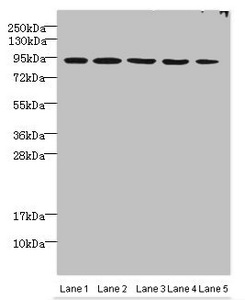
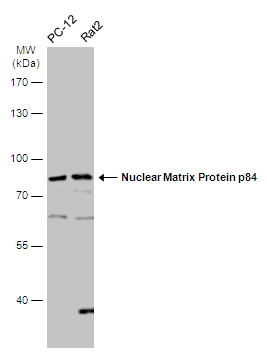
![Various whole cell extracts (30 μg) were separated by 7.5% SDS-PAGE, and the membrane was blotted with Nuclear Matrix Protein p84 antibody [C1C3] (GTX102919) diluted at 1:1000. The HRP-conjugated anti-rabbit IgG antibody (GTX213110-01) was used to detect the primary antibody.](https://www.genetex.com/upload/website/prouct_img/normal/GTX102919/GTX102919_40163_20170825_WB_R_w_23060119_665.webp "Various whole cell extracts (30 μg) were separated by 7.5% SDS-PAGE, and the membrane was blotted with Nuclear Matrix Protein p84 antibody [C1C3] (GTX102919) diluted at 1:1000. The HRP-conjugated anti-rabbit IgG antibody (GTX213110-01) was used to detect the primary antibody.")
 antibody at 1:500 dilution.
Antigen Retrieval: Trilogy? (EDTA based, pH 8.0) buffer, 15min")
Non-transfected (–) and transfected (+) 293T whole cell extracts (30 μg) were separated by 7.5% SDS-PAGE, and the membrane was blotted with Nuclear Matrix Protein p84 antibody [C1C3] (GTX102919) diluted at 1:2000. The HRP-conjugated anti-rabbit IgG antibody (GTX213110-01) was used to detect the primary antibody.
Nuclear Matrix Protein p84 antibody [C1C3]
GTX102919
ApplicationsImmunoFluorescence, ImmunoPrecipitation, Western Blot, ChIP Chromatin ImmunoPrecipitation, ImmunoCytoChemistry, ImmunoHistoChemistry, ImmunoHistoChemistry Paraffin
Product group Antibodies
ReactivityAvian, Human, Mouse, Rat
TargetTHOC1
Overview
- SupplierGeneTex
- Product NameNuclear Matrix Protein p84 antibody [C1C3]
- Delivery Days Customer9
- Application Supplier NoteWB: 1:500-1:3000. ICC/IF: 1:100-1:1000. IHC-P: 1:100-1:1000. IP: 1:100-1:500. *Optimal dilutions/concentrations should be determined by the researcher.Not tested in other applications.
- ApplicationsImmunoFluorescence, ImmunoPrecipitation, Western Blot, ChIP Chromatin ImmunoPrecipitation, ImmunoCytoChemistry, ImmunoHistoChemistry, ImmunoHistoChemistry Paraffin
- CertificationResearch Use Only
- ClonalityPolyclonal
- Concentration1 mg/ml
- ConjugateUnconjugated
- Gene ID9984
- Target nameTHOC1
- Target descriptionTHO complex subunit 1
- Target synonymsDFNA86, HPR1, P84, P84N5, THO complex subunit 1, THO complex 1, hTREX84, nuclear matrix protein p84, tho1
- HostRabbit
- IsotypeIgG
- Protein IDQ96FV9
- Protein NameTHO complex subunit 1
- Scientific DescriptionHPR1 is part of the TREX (transcription/export) complex, which includes TEX1 (MIM 606929), THO2 (MIM 300395), ALY (MIM 604171), and UAP56 (MIM 606390).[supplied by OMIM]
- ReactivityAvian, Human, Mouse, Rat
- Storage Instruction-20°C or -80°C,2°C to 8°C
- UNSPSC41116161
Datasheet
Related products
Product group Antibodies
ApplicationsImmunoFluorescence, Western Blot, ImmunoCytoChemistry, ImmunoHistoChemistry
ReactivityHuman, Mouse, Rat
- SizePrice
Product group Antibodies
Anti-THOC1 Antibody144-08179
ApplicationsImmunoFluorescence, Western Blot, ImmunoHistoChemistry
ReactivityHuman, Mouse, Rat
TargetTHOC1
- SizePrice
Product group Antibodies
ApplicationsImmunoFluorescence, Western Blot, ImmunoCytoChemistry, ImmunoHistoChemistry, ImmunoHistoChemistry Paraffin
ReactivityHuman, Mouse, Rat
TargetTHOC1
- SizePrice
Product group Antibodies
THOC1 AntibodyCSB-PA853419ESR1HU
ApplicationsWestern Blot, ChIP Chromatin ImmunoPrecipitation, ELISA, ImmunoHistoChemistry
ReactivityHuman, Mouse
TargetTHOC1
- SizePrice

Product group Antibodies
ApplicationsImmunoFluorescence, Western Blot, ImmunoCytoChemistry, ImmunoHistoChemistry
ReactivityHuman, Mouse, Rat
TargetTHOC1
- SizePrice
Product group Antibodies

p84 / THOC1 AntibodyLS-C409715
ApplicationsWestern Blot, ImmunoHistoChemistry
ReactivityHuman, Mouse, Rat
TargetTHOC1
- SizePrice
Product group Antibodies
Nuclear Matrix Protein p84 antibodyGTX118740
ApplicationsImmunoFluorescence, ImmunoPrecipitation, Western Blot, ImmunoCytoChemistry
ReactivityHuman, Mouse, Rat
TargetTHOC1
- SizePrice
![p84 antibody [5E10] (HRP) detects p84 protein by western blot analysis. A. 30 μg HeLa whole cell lysate/extract B. 30 μg HeLa nuclear lysate/extract 7.5 % SDS-PAGE p84 antibody [5E10] (HRP) (GTX670220-01) dilution: 1:1000](https://www.genetex.com/upload/website/prouct_img/normal/GTX70220-01/GTX70220-01_41333_WB_Fraction_w_23061221_806.webp)
Product group Antibodies
ApplicationsWestern Blot
ReactivityHuman, Mouse
TargetTHOC1
- SizePrice
![p84 antibody [5E10] immunoprecipitates p84 protein in IP experiments. IP Sample: HepG2 whole cell lysate/extract A : 30 μg whole cell lysate/extract of p84 protein expressing HepG2 cells B : Control with 3 μg of pre-immune mouse IgG C : Immunoprecipitation of p84 by 3 μg of p84 antibody [5E10] (GTX70220) 7.5% SDS-PAGE The immunoprecipitated p84 protein was detected by p84 antibody [5E10] (GTX70220) diluted at 1 : 1000. EasyBlot anti-rabbit IgG (HRP) (GTX221667-01) was used as a secondary reagent.](https://www.genetex.com/upload/website/prouct_img/normal/GTX70220/GTX70220_41295_IP_w_23061221_303.webp)
Product group Antibodies
ApplicationsImmunoFluorescence, ImmunoPrecipitation, Western Blot, ChIP Chromatin ImmunoPrecipitation, ImmunoCytoChemistry, ImmunoHistoChemistry, ImmunoHistoChemistry Paraffin
ReactivityHamster, Human, Monkey, Mouse, Rat
TargetTHOC1
- SizePrice
Product group Antibodies
ApplicationsImmunoFluorescence, Western Blot, ImmunoCytoChemistry, ImmunoHistoChemistry, ImmunoHistoChemistry Paraffin
ReactivityHuman, Mouse, Rat
TargetTHOC1
- SizePrice