

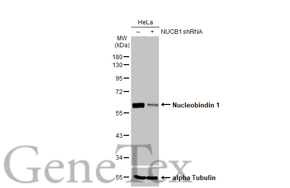
Non-transfected (–) and transfected (+) HeLa whole cell extracts (30 μg) were separated by 10% SDS-PAGE, and the membrane was blotted with Nucleobindin 1 antibody [HL2532] (GTX638901) diluted at 1:1000. The HRP-conjugated anti-rabbit IgG antibody (GTX213110-01) was used to detect the primary antibody.
![Nucleobindin 1 antibody [HL2532] detects Nucleobindin 1 protein at Golgi apparatus by immunofluorescent analysis. Sample: HeLa cells were fixed in 4% paraformaldehyde at RT for 15 min. Green: Nucleobindin 1 stained by Nucleobindin 1 antibody [HL2532] (GTX638901) diluted at 1:500. Red: alpha Tubulin, a cytoskeleton marker, stained by alpha Tubulin antibody [GT114] (GTX628802) diluted at 1:1000. Blue: Fluoroshield with DAPI (GTX30920).](https://www.genetex.com/upload/website/prouct_img/normal/GTX638901/GTX638901_T-45124_20230825_ICC_IF_23090619_837.webp "Nucleobindin 1 antibody [HL2532] detects Nucleobindin 1 protein at Golgi apparatus by immunofluorescent analysis. Sample: HeLa cells were fixed in 4% paraformaldehyde at RT for 15 min. Green: Nucleobindin 1 stained by Nucleobindin 1 antibody [HL2532] (GTX638901) diluted at 1:500. Red: alpha Tubulin, a cytoskeleton marker, stained by alpha Tubulin antibody [GT114] (GTX628802) diluted at 1:1000. Blue: Fluoroshield with DAPI (GTX30920).")
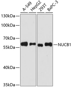
![Various whole cell extracts (30 μg) were separated by 10% SDS-PAGE, and the membrane was blotted with Nucleobindin 1 antibody [HL2532] (GTX638901) diluted at 1:1000. The HRP-conjugated anti-rabbit IgG antibody (GTX213110-01) was used to detect the primary antibody. Corresponding RNA expression data for the same cell lines are based on Human Protein Atlas program.](https://www.genetex.com/upload/website/prouct_img/normal/GTX638901/GTX638901_45194_20231027_WB_TPM_watermark_23103019_461.webp "Various whole cell extracts (30 μg) were separated by 10% SDS-PAGE, and the membrane was blotted with Nucleobindin 1 antibody [HL2532] (GTX638901) diluted at 1:1000. The HRP-conjugated anti-rabbit IgG antibody (GTX213110-01) was used to detect the primary antibody. Corresponding RNA expression data for the same cell lines are based on Human Protein Atlas program.")
![Various whole cell extracts (30 μg) were separated by 10% SDS-PAGE, and the membrane was blotted with Nucleobindin 1 antibody [HL2532] (GTX638901) diluted at 1:1000. The HRP-conjugated anti-rabbit IgG antibody (GTX213110-01) was used to detect the primary antibody.](https://www.genetex.com/upload/website/prouct_img/normal/GTX638901/GTX638901_T-45124_20231208_WB_M_R_23121122_957.webp "Various whole cell extracts (30 μg) were separated by 10% SDS-PAGE, and the membrane was blotted with Nucleobindin 1 antibody [HL2532] (GTX638901) diluted at 1:1000. The HRP-conjugated anti-rabbit IgG antibody (GTX213110-01) was used to detect the primary antibody.")
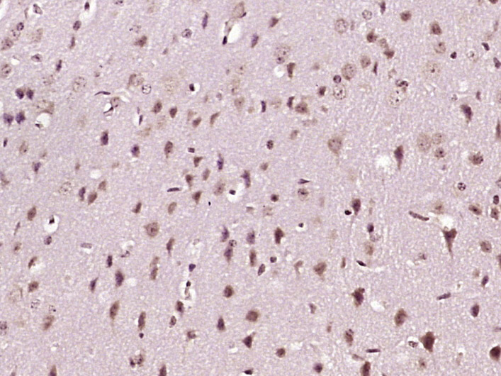
![Nucleobindin 1 antibody [HL2532] detects secreted Nucleobindin 1 protein by immunohistochemical analysis. Sample: Paraffin-embedded rat cerebellum. Nucleobindin 1 stained by Nucleobindin 1 antibody [HL2532] (GTX638901) diluted at 1:100. Antigen Retrieval: Citrate buffer, pH 6.0, 15 min](https://www.genetex.com/upload/website/prouct_img/normal/GTX638901/GTX638901_T-45124_20231208_IHC-P_R_24010223_410.webp "Nucleobindin 1 antibody [HL2532] detects secreted Nucleobindin 1 protein by immunohistochemical analysis. Sample: Paraffin-embedded rat cerebellum. Nucleobindin 1 stained by Nucleobindin 1 antibody [HL2532] (GTX638901) diluted at 1:100. Antigen Retrieval: Citrate buffer, pH 6.0, 15 min")
![Nucleobindin 1 antibody [HL2532] detects secreted Nucleobindin 1 protein by immunohistochemical analysis. Sample: Paraffin-embedded mouse cerebellum. Nucleobindin 1 stained by Nucleobindin 1 antibody [HL2532] (GTX638901) diluted at 1:100. Antigen Retrieval: Citrate buffer, pH 6.0, 15 min](https://www.genetex.com/upload/website/prouct_img/normal/GTX638901/GTX638901_T-45124_20231208_IHC-P_M_24010223_309.webp "Nucleobindin 1 antibody [HL2532] detects secreted Nucleobindin 1 protein by immunohistochemical analysis. Sample: Paraffin-embedded mouse cerebellum. Nucleobindin 1 stained by Nucleobindin 1 antibody [HL2532] (GTX638901) diluted at 1:100. Antigen Retrieval: Citrate buffer, pH 6.0, 15 min")
Non-transfected (–) and transfected (+) HeLa whole cell extracts (30 μg) were separated by 10% SDS-PAGE, and the membrane was blotted with Nucleobindin 1 antibody [HL2532] (GTX638901) diluted at 1:1000. The HRP-conjugated anti-rabbit IgG antibody (GTX213110-01) was used to detect the primary antibody.
Nucleobindin 1 antibody [HL2532]
GTX638901
ApplicationsImmunoFluorescence, Western Blot, ImmunoCytoChemistry, ImmunoHistoChemistry, ImmunoHistoChemistry Paraffin
Product group Antibodies
ReactivityHuman, Mouse, Rat
TargetNUCB1
Overview
- SupplierGeneTex
- Product NameNucleobindin 1 antibody [HL2532]
- Delivery Days Customer9
- Application Supplier NoteWB: 1:500-1:3000. *Optimal dilutions/concentrations should be determined by the researcher.Not tested in other applications.
- ApplicationsImmunoFluorescence, Western Blot, ImmunoCytoChemistry, ImmunoHistoChemistry, ImmunoHistoChemistry Paraffin
- CertificationResearch Use Only
- ClonalityMonoclonal
- Clone IDHL2532
- Concentration1 mg/ml
- ConjugateUnconjugated
- Gene ID4924
- Target nameNUCB1
- Target descriptionnucleobindin 1
- Target synonymsCALNUC, NUC, nucleobindin-1
- HostRabbit
- IsotypeIgG
- Protein IDQ02818
- Protein NameNucleobindin-1
- Scientific DescriptionThis gene encodes a member of a small calcium-binding EF-hand protein family. The encoded protein is thought to have a key role in Golgi calcium homeostasis and Ca(2+)-regulated signal transduction events. [provided by RefSeq, Jun 2010]
- ReactivityHuman, Mouse, Rat
- Storage Instruction-20°C or -80°C,2°C to 8°C
- UNSPSC41116161
Datasheet
Related products
Product group Antibodies
Anti-NUCB1 AntibodyA90694
ApplicationsImmunoFluorescence, Western Blot, ImmunoCytoChemistry, ImmunoHistoChemistry
ReactivityHuman, Mouse, Rat
- SizePrice
Product group Antibodies
ApplicationsImmunoFluorescence, Western Blot, ImmunoCytoChemistry, ImmunoHistoChemistry
ReactivityHuman, Mouse, Rat
TargetNUCB1
- SizePrice
Product group Antibodies
Anti-NUCB1 Antibody144-61376
ApplicationsWestern Blot
ReactivityHuman
TargetNUCB1
- SizePrice
Product group Antibodies

ApplicationsImmunoPrecipitation, Western Blot
ReactivityHuman
TargetNUCB1
- SizePrice
Product group Antibodies
Nucleobindin 1 Polyclonal AntibodyBS-19500R
ApplicationsImmunoFluorescence, Western Blot, ImmunoCytoChemistry, ImmunoHistoChemistry, ImmunoHistoChemistry Frozen, ImmunoHistoChemistry Paraffin
ReactivityBovine, Canine, Equine, Human, Mouse, Porcine, Rabbit, Rat, Sheep
TargetNUCB1
- SizePrice
Product group Antibodies
ApplicationsImmunoPrecipitation, Western Blot, ImmunoCytoChemistry, ImmunoHistoChemistry
ReactivityMouse, Rat
TargetNUCB1
- SizePrice
Product group Antibodies
NUCB1 AntibodyCSB-PA016145GA01HU
ApplicationsWestern Blot, ELISA, ImmunoHistoChemistry
ReactivityHuman, Mouse, Rat
TargetNUCB1
- SizePrice

Product group Antibodies
Anti-NUCB1 AntibodyHPA008176
ApplicationsWestern Blot, ImmunoCytoChemistry, ImmunoHistoChemistry
ReactivityHuman
TargetNUCB1
- SizePrice
![Nucleobindin 1 antibody [N3C3] detects Nucleobindin 1 protein at cytosol on mouse kidney by immunohistochemical analysis. Sample: Paraffin-embedded mouse kidney. Nucleobindin 1 antibody [N3C3] (GTX114593) dilution: 1:500.
Antigen Retrieval: Trilogy? (EDTA based, pH 8.0) buffer, 15min](https://www.genetex.com/upload/website/prouct_img/normal/GTX114593/GTX114593_40205_IHC_M_w_23060518_768.webp)
Product group Antibodies
Nucleobindin 1 antibody [N3C3]GTX114593
ApplicationsImmunoFluorescence, Western Blot, ImmunoCytoChemistry, ImmunoHistoChemistry, ImmunoHistoChemistry Paraffin
ReactivityHuman, Mouse
TargetNUCB1
- SizePrice
Product group Antibodies
ApplicationsImmunoFluorescence, Western Blot, ELISA, ImmunoCytoChemistry
ReactivityHuman
TargetNUCB1
- SizePrice