

![Nucleolin(364-5), Biotin conjugate, 0.1mg/mL [26628-22-8]](https://biotium.com/wp-content/uploads/2016/12/CF555-nucleolin-CF488A-phalloidin-Hoecsht-HeLa-b.jpg "Nucleolin(364-5), Biotin conjugate, 0.1mg/mL [26628-22-8]")
![Nucleolin(364-5), Biotin conjugate, 0.1mg/mL [26628-22-8]](https://biotium.com/wp-content/uploads/2016/12/BNUB0527-1-1.jpg "Nucleolin(364-5), Biotin conjugate, 0.1mg/mL [26628-22-8]")
![Nucleolin(364-5), Biotin conjugate, 0.1mg/mL [26628-22-8]](https://biotium.com/wp-content/uploads/2016/12/CellBrite-Fix-640-CF488A-Nucleolin-364-5.jpg "Nucleolin(364-5), Biotin conjugate, 0.1mg/mL [26628-22-8]")
![Nucleolin(364-5), Biotin conjugate, 0.1mg/mL [26628-22-8]](https://biotium.com/wp-content/uploads/2016/12/BNUB0527-2-1.jpg "Nucleolin(364-5), Biotin conjugate, 0.1mg/mL [26628-22-8]")
![Nucleolin(364-5), Biotin conjugate, 0.1mg/mL [26628-22-8]](https://biotium.com/wp-content/uploads/2016/12/CellBrite-Blue-multicolor-IF.jpg "Nucleolin(364-5), Biotin conjugate, 0.1mg/mL [26628-22-8]")
![Nucleolin(364-5), Biotin conjugate, 0.1mg/mL [26628-22-8]](https://biotium.com/wp-content/uploads/2016/12/BNUB0527-0-1.jpg "Nucleolin(364-5), Biotin conjugate, 0.1mg/mL [26628-22-8]")
![Nucleolin(364-5), Biotin conjugate, 0.1mg/mL [26628-22-8]](https://biotium.com/wp-content/uploads/2016/12/BNUB0527-3-1.jpg "Nucleolin(364-5), Biotin conjugate, 0.1mg/mL [26628-22-8]")
![Nucleolin(364-5), Biotin conjugate, 0.1mg/mL [26628-22-8]](https://biotium.com/wp-content/uploads/2016/12/CF647-nucleolin-IgG1-isotype-flow.jpg "Nucleolin(364-5), Biotin conjugate, 0.1mg/mL [26628-22-8]")
![Nucleolin(364-5), Biotin conjugate, 0.1mg/mL [26628-22-8]](https://biotium.com/wp-content/uploads/2016/12/CF640R-nucleolin-Cy5-WB-markers-2.jpg "Nucleolin(364-5), Biotin conjugate, 0.1mg/mL [26628-22-8]")
Nucleolin(364-5), Biotin conjugate, 0.1mg/mL [26628-22-8]
BNCB0527
ApplicationsFlow Cytometry, ImmunoFluorescence, Western Blot, ImmunoHistoChemistry, ImmunoHistoChemistry Paraffin
Product group Antibodies
ReactivityBovine, Human, Mouse
TargetNCL
Overview
- SupplierBiotium
- Product NameNucleolin(364-5), Biotin conjugate, 0.1mg/mL [26628-22-8]
- Delivery Days Customer9
- ApplicationsFlow Cytometry, ImmunoFluorescence, Western Blot, ImmunoHistoChemistry, ImmunoHistoChemistry Paraffin
- CAS Number26628-22-8
- CertificationResearch Use Only
- ClonalityMonoclonal
- Clone ID364-5
- Concentration0.1 mg/ml
- ConjugateBiotin
- Gene ID4691
- Target nameNCL
- Target descriptionnucleolin
- Target synonymsC23, Nsr1, nucleolin
- HostMouse
- IsotypeIgG1
- Protein IDP19338
- Protein NameNucleolin
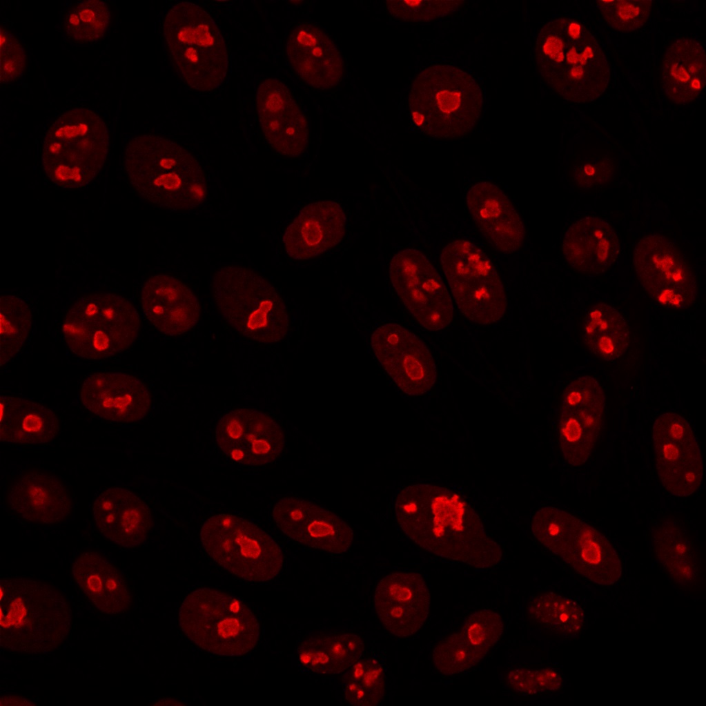
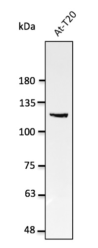
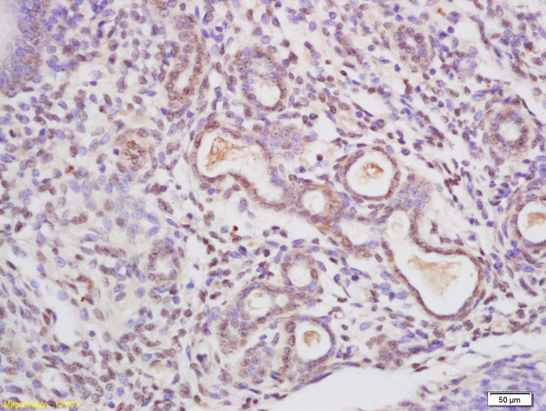
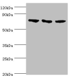
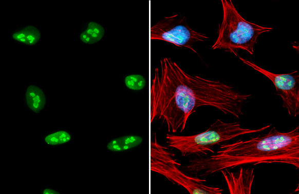
- Scientific DescriptionRecognizes Nucleolin (NCL), the major nucleolar phosphoprotein of growing eukaryotic cells. NCL is located mainly in dense fibrillar regions of the nucleolus. It is found associated with intranucleolar chromatin and pre-ribosomal particles. Human NCL gene consists of 14 exons with 13 introns and spans approximately 11 kb. It has a predicted molecular weight of ~76 kDa with an apparent MW of 100-110 kDa, attributed to phosphorylation of its N-terminal domain. It induces chromatin decondensation by binding to histone H1. It is thought to play a role in pre-rRNA transcription and ribosome assembly.This MAb can be used to stain the nucleoli in cell or tissue preparations and can be used as a marker of the nucleoli in subcellular fractions. It produces a speckled pattern in the nuclei of cells of normal and malignant cells and may be used to stain the nucleoli of cells in fixed or frozen tissue sections. It can be used with paraformaldehyde- or methanol-fixed frozen tissue or cell preparations and formalin fixed, paraffin-embedded tissue sections. Primary antibodies are available purified, or with a selection of fluorescent CF® Dyes and other labels. CF® Dyes offer exceptional brightness and photostability. Note: Conjugates of blue fluorescent dyes like CF®405S and CF®405M are not recommended for detecting low abundance targets, because blue dyes have lower fluorescence and can give higher non-specific background than other dye colors.
- SourceAnimal
- ReactivityBovine, Human, Mouse
- Storage Instruction2°C to 8°C,RT
- UNSPSC41116161
MSDS
Related products
Product group Antibodies
Anti-Nucleolin AntibodyA121665
ApplicationsWestern Blot
ReactivityCanine, Human, Monkey, Mouse, Rat
- SizePrice
Product group Antibodies
Anti-NCL Antibody144-05904
ApplicationsWestern Blot, ImmunoHistoChemistry
ReactivityHuman, Mouse, Rat
TargetNCL
- SizePrice

Product group Antibodies
Anti-Nucleolin/NCL Antibody Picoband(r)A00228-1-CARRIER-FREE
ApplicationsFlow Cytometry, ImmunoFluorescence, Western Blot, ELISA, ImmunoCytoChemistry, ImmunoHistoChemistry
ReactivityHuman, Monkey, Mouse, Rat
TargetNCL
- SizePrice
Product group Antibodies
ApplicationsFlow Cytometry, ImmunoFluorescence, ELISA, ImmunoCytoChemistry, ImmunoHistoChemistry, ImmunoHistoChemistry Frozen, ImmunoHistoChemistry Paraffin
ReactivityBovine, Canine, Equine, Human, Mouse, Porcine, Rat
TargetNCL
- SizePrice
Product group Antibodies
ApplicationsImmunoPrecipitation, Western Blot, ImmunoCytoChemistry, ImmunoHistoChemistry
ReactivityMouse
TargetNCL
- SizePrice
Product group Antibodies
NCL AntibodyCSB-PA015535DSR1HU
ApplicationsWestern Blot, ELISA
ReactivityHuman
TargetNCL
- SizePrice
Product group Antibodies

NCL / Nucleolin AntibodyLS-C408894
ApplicationsWestern Blot, ImmunoHistoChemistry
ReactivityHuman, Mouse, Rat
TargetNCL
- SizePrice
Product group Antibodies
Nucleolin antibodyGTX132778
ApplicationsImmunoFluorescence, Western Blot, ImmunoCytoChemistry
ReactivityHuman
TargetNCL
- SizePrice

Product group Antibodies
Anti-NCL AntibodyHPA023981
ApplicationsChIP Chromatin ImmunoPrecipitation, ImmunoCytoChemistry, ImmunoHistoChemistry
ReactivityHuman
TargetNCL
- SizePrice