

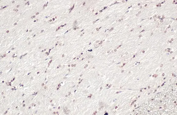
OLIG2 antibody detects OLIG2 protein at nucleus by immunohistochemical analysis. Sample: Paraffin-embedded rat brain. OLIG2 stained by OLIG2 antibody (GTX132732) diluted at 1:500. Antigen Retrieval: Citrate buffer, pH 6.0, 15 min
![OLIG2 antibody detects OLIG2 protein by immunohistochemical analysis. Sample: Paraffin-embedded mouse E10.5 embryo. Green: OLIG2 stained by OLIG2 antibody (GTX132732) diluted at 1:500. Red: beta Tubulin 3/ Tuj1, a Cytoskeleton marker, stained by beta Tubulin 3/ Tuj1 antibody [GT11710] (GTX631836) diluted at 1:500. Blue: Fluoroshield with DAPI (GTX30920). Antigen Retrieval: Citrate buffer, pH 6.0, 15 min](https://www.genetex.com/upload/website/prouct_img/normal/GTX132732/GTX132732_44923_20230217_IHC-P_M_23021401_367.webp "OLIG2 antibody detects OLIG2 protein by immunohistochemical analysis. Sample: Paraffin-embedded mouse E10.5 embryo. Green: OLIG2 stained by OLIG2 antibody (GTX132732) diluted at 1:500. Red: beta Tubulin 3/ Tuj1, a Cytoskeleton marker, stained by beta Tubulin 3/ Tuj1 antibody [GT11710] (GTX631836) diluted at 1:500. Blue: Fluoroshield with DAPI (GTX30920). Antigen Retrieval: Citrate buffer, pH 6.0, 15 min")
![OLIG2 antibody detects OLIG2 protein by immunohistochemical analysis. Sample: Paraffin-embedded mouse E10.5 embryo. Green: OLIG2 stained by OLIG2 antibody (GTX132732) diluted at 1:500. Red: beta Tubulin 3/ Tuj1, a Cytoskeleton marker, stained by beta Tubulin 3/ Tuj1 antibody [GT11710] (GTX631836) diluted at 1:500. Blue: Fluoroshield with DAPI (GTX30920). Antigen Retrieval: Citrate buffer, pH 6.0, 15 min](https://www.genetex.com/upload/website/prouct_img/normal/GTX132732/GTX132732_44923_20230217_IHC-P_M_1_23021401_732.webp "OLIG2 antibody detects OLIG2 protein by immunohistochemical analysis. Sample: Paraffin-embedded mouse E10.5 embryo. Green: OLIG2 stained by OLIG2 antibody (GTX132732) diluted at 1:500. Red: beta Tubulin 3/ Tuj1, a Cytoskeleton marker, stained by beta Tubulin 3/ Tuj1 antibody [GT11710] (GTX631836) diluted at 1:500. Blue: Fluoroshield with DAPI (GTX30920). Antigen Retrieval: Citrate buffer, pH 6.0, 15 min")
![OLIG2 antibody detects OLIG2 protein by immunofluorescent analysis. Sample: DIV9 rat E18 primary cortical neurons and glia cells were fixed in 4% paraformaldehyde at RT for 15 min. Green: OLIG2 protein stained by OLIG2 antibody (GTX132732) diluted at 1:500. Red: beta Tubulin 3/ Tuj1, stained by beta Tubulin 3/ Tuj1 antibody [GT886] (GTX631830) diluted at 1:500. Blue: Fluoroshield with DAPI (GTX30920).](https://www.genetex.com/upload/website/prouct_img/normal/GTX132732/GTX132732_42774_20170705_IFA_R_w_23060523_172.webp "OLIG2 antibody detects OLIG2 protein by immunofluorescent analysis. Sample: DIV9 rat E18 primary cortical neurons and glia cells were fixed in 4% paraformaldehyde at RT for 15 min. Green: OLIG2 protein stained by OLIG2 antibody (GTX132732) diluted at 1:500. Red: beta Tubulin 3/ Tuj1, stained by beta Tubulin 3/ Tuj1 antibody [GT886] (GTX631830) diluted at 1:500. Blue: Fluoroshield with DAPI (GTX30920).")
 diluted at 1:5000. Antigen Retrieval: Tris-EDTA buffer, pH 9.0, 15 min")
OLIG2 antibody detects OLIG2 protein at nucleus by immunohistochemical analysis. Sample: Paraffin-embedded rat brain. OLIG2 stained by OLIG2 antibody (GTX132732) diluted at 1:500. Antigen Retrieval: Citrate buffer, pH 6.0, 15 min
OLIG2 antibody
GTX132732
ApplicationsImmunoFluorescence, Western Blot, ImmunoCytoChemistry, ImmunoHistoChemistry, ImmunoHistoChemistry Frozen, ImmunoHistoChemistry Paraffin
Product group Antibodies
ReactivityHuman, Mouse, Porcine, Rat
TargetOLIG2
Overview
- SupplierGeneTex
- Product NameOLIG2 antibody
- Delivery Days Customer9
- Application Supplier NoteWB: 1:500-1:3000. ICC/IF: 1:100-1:1000. *Optimal dilutions/concentrations should be determined by the researcher.Not tested in other applications.
- ApplicationsImmunoFluorescence, Western Blot, ImmunoCytoChemistry, ImmunoHistoChemistry, ImmunoHistoChemistry Frozen, ImmunoHistoChemistry Paraffin
- CertificationResearch Use Only
- ClonalityPolyclonal
- Concentration1 mg/ml
- ConjugateUnconjugated
- Gene ID10215
- Target nameOLIG2
- Target descriptionoligodendrocyte transcription factor 2
- Target synonymsBHLHB1, OLIGO2, PRKCBP2, RACK17, bHLHe19, oligodendrocyte transcription factor 2, basic domain, helix-loop-helix protein, class B, 1, class B basic helix-loop-helix protein 1, class E basic helix-loop-helix protein 19, human protein kinase C-binding protein RACK17, oligodendrocyte lineage transcription factor 2, oligodendrocyte-specific bHLH transcription factor 2, protein kinase C-binding protein 2
- HostRabbit
- IsotypeIgG
- Protein IDQ13516
- Protein NameOligodendrocyte transcription factor 2
- Scientific DescriptionThis gene encodes a basic helix-loop-helix transcription factor which is expressed in oligodendroglial tumors of the brain. The protein is an essential regulator of ventral neuroectodermal progenitor cell fate. The gene is involved in a chromosomal translocation t(14;21)(q11.2;q22) associated with T-cell acute lymphoblastic leukemia. Its chromosomal location is within a region of chromosome 21 which has been suggested to play a role in learning deficits associated with Down syndrome. [provided by RefSeq]
- ReactivityHuman, Mouse, Porcine, Rat
- Storage Instruction-20°C or -80°C,2°C to 8°C
- UNSPSC41116161
Datasheet
Related products
Product group Antibodies
Anti-OLIG2 Antibody144-62495
ApplicationsWestern Blot, ImmunoHistoChemistry
ReactivityHuman, Mouse, Rat
TargetOLIG2
- SizePrice
Product group Antibodies

OLIG2 AntibodyLS-C761202
ApplicationsWestern Blot
ReactivityHuman, Mouse, Rat
TargetOLIG2
- SizePrice

Product group Antibodies
Anti-OLIG2 Antibody Picoband(r)A02247-2-CARRIER-FREE
ApplicationsWestern Blot
ReactivityHuman, Mouse, Rat
TargetOLIG2
- SizePrice
Product group Antibodies
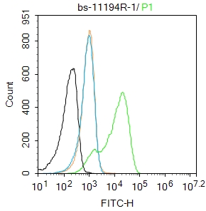
OLIG2 Polyclonal Antibodybs-11194R
ApplicationsFlow Cytometry, ImmunoFluorescence, ELISA, ImmunoCytoChemistry, ImmunoHistoChemistry, ImmunoHistoChemistry Frozen, ImmunoHistoChemistry Paraffin
ReactivityBovine, Canine, Chicken, Human, Mouse, Porcine, Rabbit, Rat, Sheep
- SizePrice
Product group Antibodies
ApplicationsImmunoPrecipitation, Western Blot, ImmunoCytoChemistry, ImmunoHistoChemistry
TargetOLIG2
- SizePrice
Product group Antibodies
OLIG2 AntibodyCSB-PA016329GA01HU
ApplicationsWestern Blot, ELISA, ImmunoHistoChemistry
ReactivityHuman, Mouse, Rat
TargetOLIG2
- SizePrice
Product group Antibodies
References
OLIG2 antibodyGTX31569
ApplicationsWestern Blot, ELISA, ImmunoHistoChemistry, ImmunoHistoChemistry Paraffin
ReactivityHuman, Mouse, Rat
TargetOLIG2
- SizePrice

Product group Antibodies
Anti-OLIG2 AntibodyHPA003254
ApplicationsWestern Blot, ChIP Chromatin ImmunoPrecipitation, ImmunoHistoChemistry
ReactivityHuman
TargetOLIG2
- SizePrice
![WB analysis of HEK293 (1) and OLIG2(AA: 1-122)-hIgGFc transfected HEK293 (2) cell lysate using GTX60398 OLIG2 antibody [1G11].](https://www.genetex.com/upload/website/prouct_img/normal/GTX60398/GTX60398_20170912_WB_w_23061123_784.webp)
Product group Antibodies
OLIG2 antibody [1G11]GTX60398
ApplicationsImmunoFluorescence, Western Blot, ELISA, ImmunoCytoChemistry
ReactivityHuman
TargetOLIG2
- SizePrice
![OLIG2 antibody [HL1072] detects OLIG2 protein at nucleus by immunohistochemical analysis. Sample: Paraffin-embedded mouse brain. OLIG2 stained by OLIG2 antibody [HL1072] (GTX636104) diluted at 1:100. Antigen Retrieval: Citrate buffer, pH 6.0, 15 min](https://www.genetex.com/upload/website/prouct_img/normal/GTX636104/GTX636104_45040_20230526_IHC-P_M_23060622_235.webp)
Product group Antibodies
OLIG2 antibody [HL1072]GTX636104
ApplicationsImmunoFluorescence, ImmunoCytoChemistry, ImmunoHistoChemistry, ImmunoHistoChemistry Frozen, ImmunoHistoChemistry Paraffin
ReactivityHuman, Mouse, Rat
TargetOLIG2
- SizePrice