

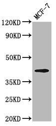
Western Blot Positive WB detected in: MCF-7 whole cell lysate All lanes: OPN1MW antibody at 3.2microg/ml Secondary Goat polyclonal to rabbit IgG at 1/50000 dilution Predicted band size: 41 kDa Observed band size: 41 kDa
.")
Western Blot Positive WB detected in: MCF-7 whole cell lysate All lanes: OPN1MW antibody at 3.2microg/ml Secondary Goat polyclonal to rabbit IgG at 1/50000 dilution Predicted band size: 41 kDa Observed band size: 41 kDa
OPN1MW Antibody
CSB-PA016352LA01HU
ApplicationsImmunoFluorescence, Western Blot, ELISA
Product group Antibodies
ReactivityHuman
TargetOPN1MW
Overview
- SupplierCusabio
- Product NameOPN1MW Antibody
- Delivery Days Customer20
- ApplicationsImmunoFluorescence, Western Blot, ELISA
- CertificationResearch Use Only
- ClonalityPolyclonal
- ConjugateUnconjugated
- Gene ID2652
- Target nameOPN1MW
- Target descriptionopsin 1, medium wave sensitive
- Target synonymsCBBM, CBD, COD5, GCP, GOP, OPN1MW1, medium-wave-sensitive opsin 1, Medium-wave-sensitive opsin 2, Opsin 1 cone pigments medium-wave-sensitive 2, cone dystrophy 5 (X-linked), green cone photoreceptor pigment, green cone pigment, green-sensitive opsin, opsin 1 (cone pigments), medium-wave-sensitive, photopigment apoprotein
- HostRabbit
- IsotypeIgG
- Protein IDP04001
- Protein NameMedium-wave-sensitive opsin 1
- Scientific DescriptionVisual pigments are the light-absorbing molecules that mediate vision. They consist of an apoprotein, opsin, covalently linked to cis-retinal.
- ReactivityHuman
- Storage Instruction-20°C or -80°C
- UNSPSC41116161
Related products
Product group Antibodies
ApplicationsWestern Blot, ELISA
ReactivityHuman, Mouse, Rat
TargetOPN1MW
- SizePrice
Product group Antibodies
Opn1Mw Polyclonal AntibodyCAC11491
ApplicationsImmunoFluorescence, Western Blot, ELISA
TargetOPN1MW
- SizePrice
Product group Antibodies
OPN1MW AntibodyPACO59824
ApplicationsImmunoFluorescence, Western Blot, ELISA
ReactivityHuman
TargetOPN1MW
- SizePrice
![Non-transfected (–) and transfected (+) boiled and unboiled 293T whole cell extracts (30 μg) were separated by 10% SDS-PAGE, and the membrane was blotted with OPN1MW2 antibody [HL3435] (GTX641277) diluted at 1:3000. The HRP-conjugated anti-rabbit IgG antibody (GTX213110-01) was used to detect the primary antibody.](https://www.genetex.com/upload/website/prouct_img/normal/GTX641277/GTX641277_T-45586_20241115_WB_B_ub_24112622_559.webp)
Product group Antibodies
Opsin Red / Green antibody [HL3435]GTX641277
ApplicationsImmunoFluorescence, Western Blot, ImmunoCytoChemistry, ImmunoHistoChemistry, ImmunoHistoChemistry Frozen, ImmunoHistoChemistry Paraffin
ReactivityHuman, Mouse, Rat
TargetOPN1MW
- SizePrice
Product group Antibodies

OPN1MW / GCP Antibody (HRP)LS-C680982
ApplicationsELISA
ReactivityHuman
TargetOPN1MW
- SizePrice