

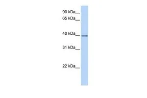
WB analysis of human fetal heart tissue using GTX45339 OPTC antibody at 0.2-1μg/ml.
WB analysis of human fetal heart tissue using GTX45339 OPTC antibody at 0.2-1μg/ml.
OPTC antibody, C-term
GTX45339
ApplicationsWestern Blot
Product group Antibodies
ReactivityHuman
TargetOPTC
Overview
- SupplierGeneTex
- Product NameOPTC antibody, C-term
- Delivery Days Customer9
- Application Supplier NoteWB: 0.2-2.5 ug/ml. *Optimal dilutions/concentrations should be determined by the researcher.Not tested in other applications.
- ApplicationsWestern Blot
- CertificationResearch Use Only
- ClonalityPolyclonal
- Concentration0.5-1 mg/ml
- ConjugateUnconjugated
- Gene ID26254
- Target nameOPTC
- Target descriptionopticin
- Target synonymsOPT, opticin, oculoglycan
- HostRabbit
- IsotypeIgG
- Protein IDQ9UBM4
- Protein NameOpticin
- Scientific DescriptionOpticin belongs to class III of the small leucine-rich repeat protein (SLRP) family. Members of this family are typically associated with the extracellular matrix. Opticin is present in significant quantities in the vitreous of the eye and also localizes to the cornea, iris, ciliary body, optic nerve, choroid, retina, and fetal liver. Opticin may noncovalently bind collagen fibrils and regulate fibril morphology, spacing, and organization. The opticin gene is mapped to a region of chromosome 1 that is associated with the inherited eye diseases age-related macular degeneration (AMD) and posterior column ataxia with retinosa pigmentosa (AXPC1). [provided by RefSeq, Jul 2008]
- ReactivityHuman
- Storage Instruction-20°C or -80°C,2°C to 8°C
- UNSPSC12352203
Datasheet
Related products

Product group Antibodies
Anti-OPTC Antibody Picoband(r)A09717-1-CARRIER-FREE
ApplicationsWestern Blot, ELISA
ReactivityHuman, Monkey
TargetOPTC
- SizePrice
Product group Antibodies
Anti-OPTC Antibody144-64450
ApplicationsWestern Blot
ReactivityHuman, Mouse, Rat
TargetOPTC
- SizePrice
Product group Antibodies
OPTC AntibodyCSB-PA016362LA01HU
ApplicationsELISA, ImmunoHistoChemistry
ReactivityHuman
TargetOPTC
- SizePrice

Product group Antibodies
Anti-OPTC AntibodyHPA034951
ApplicationsImmunoHistoChemistry
ReactivityHuman
TargetOPTC
- SizePrice
Product group Antibodies

OPTC / Opticin Antibody (C-Terminus)LS-C353384
ApplicationsWestern Blot
ReactivityHuman, Mouse, Porcine, Rat
TargetOPTC
- SizePrice
Product group Antibodies
Anti-Opticin AntibodyA28518
ApplicationsWestern Blot
ReactivityHuman, Mouse, Rat
- SizePrice