

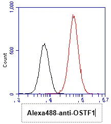
FACS analysis of HeLa cells using GTX57564 OSTF1 antibody. Cell Number: 1 x 10? cells Primary antibody: Red line Antibody amount: 2-5 μg

 transfected 293T cell lysate Loading : 10 μg Dilution : 1:1000")


FACS analysis of HeLa cells using GTX57564 OSTF1 antibody. Cell Number: 1 x 10? cells Primary antibody: Red line Antibody amount: 2-5 μg
OSTF1 antibody [AT9G4]
GTX57564
ApplicationsFlow Cytometry, ImmunoFluorescence, Western Blot, ImmunoCytoChemistry
Product group Antibodies
ReactivityHuman
TargetOSTF1
Overview
- SupplierGeneTex
- Product NameOSTF1 antibody [AT9G4]
- Delivery Days Customer9
- ApplicationsFlow Cytometry, ImmunoFluorescence, Western Blot, ImmunoCytoChemistry
- CertificationResearch Use Only
- ClonalityMonoclonal
- Clone IDAT9G4
- Concentration1 mg/ml
- ConjugateUnconjugated
- Gene ID26578
- Target nameOSTF1
- Target descriptionosteoclast stimulating factor 1
- Target synonymsOSF, SH3P2, bA235O14.1, osteoclast-stimulating factor 1
- HostMouse
- IsotypeIgG2a
- Protein IDQ92882
- Protein NameOsteoclast-stimulating factor 1
- Scientific DescriptionOsteoclast-stimulating factor-1 is an intracellular protein produced by osteoclasts that indirectly induces osteoclast formation and bone resorption (Reddy et al., 1998 [PubMed 10092216]).[supplied by OMIM, Mar 2008]
- ReactivityHuman
- Storage Instruction-20°C or -80°C,2°C to 8°C
- UNSPSC41116161
Datasheet
Related products
Product group Antibodies
OSTF1 AntibodyCSB-PA017266GA01HU
ApplicationsELISA, ImmunoHistoChemistry
ReactivityHuman, Mouse, Rat
TargetOSTF1
- SizePrice

Product group Antibodies
Anti-OSTF1 Antibody Picoband(r)A05988-1-CARRIER-FREE
ApplicationsFlow Cytometry, ImmunoFluorescence, Western Blot, ELISA, ImmunoCytoChemistry
ReactivityHuman, Mouse, Rat
TargetOSTF1
- SizePrice
Product group Antibodies

OSTF1 / OSF AntibodyLS-C747343
ApplicationsWestern Blot
ReactivityHuman, Mouse, Rat
TargetOSTF1
- SizePrice

Product group Antibodies
Anti-OSTF1 AntibodyHPA020514
ApplicationsWestern Blot, ImmunoCytoChemistry, ImmunoHistoChemistry
ReactivityHuman
TargetOSTF1
- SizePrice
Product group Antibodies
ApplicationsImmunoPrecipitation, Western Blot, ImmunoCytoChemistry, ImmunoHistoChemistry
TargetOSTF1
- SizePrice