

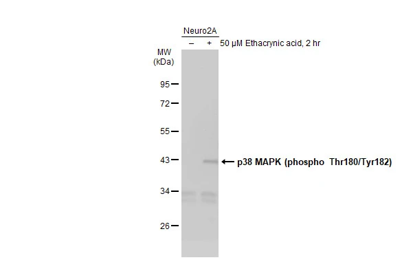
Whole cell extract (30 μg) was separated by 10% SDS-PAGE, and the membrane was blotted with p38 MAPK antibody (GTX110720) diluted at 1:500. The HRP-conjugated anti-rabbit IgG antibody (GTX213110-01) was used to detect the primary antibody.
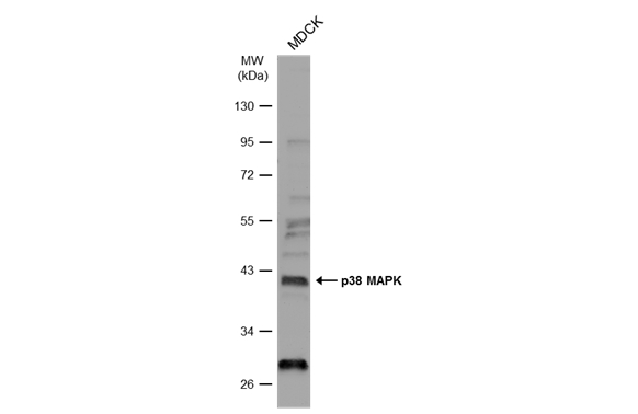
![Various whole cell extracts (30 μg) were separated by 10% SDS-PAGE, and the membrane was blotted with p38 MAPK antibody [N1C3-2] (GTX110720) diluted at 1:1000. The HRP-conjugated anti-rabbit IgG antibody (GTX213110-01) was used to detect the primary antibody.](https://www.genetex.com/upload/website/prouct_img/normal/GTX110720/GTX110720_43880_20200320_WB_w_23060500_988.webp "Various whole cell extracts (30 μg) were separated by 10% SDS-PAGE, and the membrane was blotted with p38 MAPK antibody [N1C3-2] (GTX110720) diluted at 1:1000. The HRP-conjugated anti-rabbit IgG antibody (GTX213110-01) was used to detect the primary antibody.")
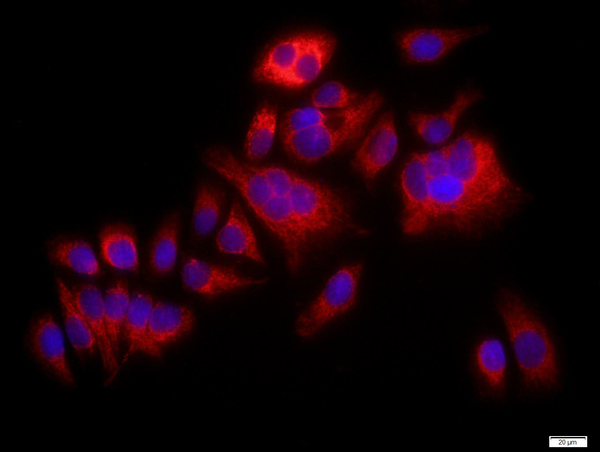
![p38 MAPK antibody [N1C3-2] detects p38 MAPK protein at cytoplasm and nucleus by immunofluorescent analysis. Sample: HeLa cells were fixed in 4% paraformaldehyde for 10 min. Green: p38 MAPK protein stained by p38 MAPK antibody [N1C3-2] (GTX110720) diluted at 1:100. Blue: Hoechst 33342 staining. Scale bar = 10 μm.](https://www.genetex.com/upload/website/prouct_img/normal/GTX110720/GTX110720_40100_20151029_IFA_w_23060500_967.webp "p38 MAPK antibody [N1C3-2] detects p38 MAPK protein at cytoplasm and nucleus by immunofluorescent analysis. Sample: HeLa cells were fixed in 4% paraformaldehyde for 10 min. Green: p38 MAPK protein stained by p38 MAPK antibody [N1C3-2] (GTX110720) diluted at 1:100. Blue: Hoechst 33342 staining. Scale bar = 10 μm.")
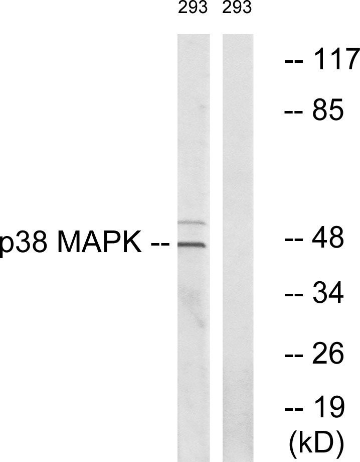
![Wild-type (WT) and p38 MAPK knockout (KO) 293T cell extracts (30 μg) were separated by 10% SDS-PAGE, and the membrane was blotted with p38 MAPK antibody [N1C3-2] (GTX110720) diluted at 1:500. The HRP-conjugated anti-rabbit IgG antibody (GTX213110-01) was used to detect the primary antibody.](https://www.genetex.com/upload/website/prouct_img/normal/GTX110720/GTX110720_40100_20180316_WB_KO_watermark_w_23060500_778.webp "Wild-type (WT) and p38 MAPK knockout (KO) 293T cell extracts (30 μg) were separated by 10% SDS-PAGE, and the membrane was blotted with p38 MAPK antibody [N1C3-2] (GTX110720) diluted at 1:500. The HRP-conjugated anti-rabbit IgG antibody (GTX213110-01) was used to detect the primary antibody.")
![p38 MAPK antibody [N1C3-2] detects p38 MAPK protein at cytoplasm in rat brain by immunohistochemical analysis. Sample: Paraffin-embedded rat brain. p38 MAPK antibody [N1C3-2] (GTX110720) diluted at 1:500.
Antigen Retrieval: Citrate buffer, pH 6.0, 15 min](https://www.genetex.com/upload/website/prouct_img/normal/GTX110720/GTX110720_40100_20160201_IHC-P_R_w_23060500_528.webp "p38 MAPK antibody [N1C3-2] detects p38 MAPK protein at cytoplasm in rat brain by immunohistochemical analysis. Sample: Paraffin-embedded rat brain. p38 MAPK antibody [N1C3-2] (GTX110720) diluted at 1:500.
Antigen Retrieval: Citrate buffer, pH 6.0, 15 min")
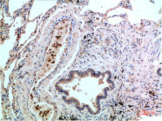
![p38 antibody [N1C3-2] detects p38 protein at nucleus on human lung adenocarcinoma by immunohistochemical analysis. Sample: Paraffin-embedded human lung adenocarcinoma. p38 antibody [N1C3-2] (GTX110720) diluted at 1:250.
Antigen Retrieval: Citrate buffer, pH 6.0, 15 min](https://www.genetex.com/upload/website/prouct_img/normal/GTX110720/GTX110720_40100_20141128_IHC_w_23060500_929.webp "p38 antibody [N1C3-2] detects p38 protein at nucleus on human lung adenocarcinoma by immunohistochemical analysis. Sample: Paraffin-embedded human lung adenocarcinoma. p38 antibody [N1C3-2] (GTX110720) diluted at 1:250.
Antigen Retrieval: Citrate buffer, pH 6.0, 15 min")


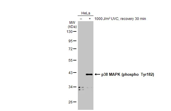
Whole cell extract (30 μg) was separated by 10% SDS-PAGE, and the membrane was blotted with p38 MAPK antibody (GTX110720) diluted at 1:500. The HRP-conjugated anti-rabbit IgG antibody (GTX213110-01) was used to detect the primary antibody.
p38 MAPK antibody [N1C3-2]
GTX110720
ApplicationsImmunoFluorescence, Western Blot, ImmunoCytoChemistry, ImmunoHistoChemistry, ImmunoHistoChemistry Paraffin
Product group Antibodies
ReactivityCanine, Human, Mouse, Rat
TargetMAPK14
Overview
- SupplierGeneTex
- Product Namep38 MAPK antibody [N1C3-2]
- Delivery Days Customer9
- Application Supplier NoteWB: 1:500-1:3000. ICC/IF: 1:100-1:1000. IHC-P: 1:100-1:1000. *Optimal dilutions/concentrations should be determined by the researcher.Not tested in other applications.
- ApplicationsImmunoFluorescence, Western Blot, ImmunoCytoChemistry, ImmunoHistoChemistry, ImmunoHistoChemistry Paraffin
- CertificationResearch Use Only
- ClonalityPolyclonal
- Concentration0.44 mg/ml
- ConjugateUnconjugated
- Gene ID1432
- Target nameMAPK14
- Target descriptionmitogen-activated protein kinase 14
- Target synonymsCSBP, CSBP1, CSBP2, CSPB1, EXIP, Mxi2, PRKM14, PRKM15, RK, SAPK2A, p38, p38ALPHA, mitogen-activated protein kinase 14, CSAID-binding protein, MAP kinase 14, MAP kinase Mxi2, MAP kinase p38 alpha, MAX-interacting protein 2, cytokine suppressive anti-inflammatory drug binding protein, mitogen-activated protein kinase p38 alpha, p38 MAP kinase, p38 mitogen activated protein kinase, p38alpha Exip, stress-activated protein kinase 2A
- HostRabbit
- IsotypeIgG
- Protein IDQ16539
- Protein NameMitogen-activated protein kinase 14
- Scientific DescriptionThe protein encoded by this gene is a member of the MAP kinase family. MAP kinases act as an integration point for multiple biochemical signals, and are involved in a wide variety of cellular processes such as proliferation, differentiation, transcription regulation and development. This kinase is activated by various environmental stresses and proinflammatory cytokines. The activation requires its phosphorylation by MAP kinase kinases (MKKs), or its autophosphorylation triggered by the interaction of MAP3K7IP1/TAB1 protein with this kinase. The substrates of this kinase include transcription regulator ATF2, MEF2C, and MAX, cell cycle regulator CDC25B, and tumor suppressor p53, which suggest the roles of this kinase in stress related transcription and cell cycle regulation, as well as in genotoxic stress response. Four alternatively spliced transcript variants of this gene encoding distinct isoforms have been reported. [provided by RefSeq]
- ReactivityCanine, Human, Mouse, Rat
- Storage Instruction-20°C or -80°C,2°C to 8°C
- UNSPSC41116161
Datasheet
Related products
Product group Antibodies
Anti-p38 MAPK AntibodyA94875
ApplicationsImmunoFluorescence, Western Blot, ELISA, ImmunoHistoChemistry
ReactivityHuman, Mouse, Rat
- SizePrice

Product group Antibodies
Anti-p38 alpha/MAPK14 Antibody Picoband(r)A00176-2-CARRIER-FREE
ApplicationsFlow Cytometry, ImmunoPrecipitation, Western Blot, ImmunoHistoChemistry
ReactivityHuman, Mouse, Rat
TargetMAPK14
- SizePrice
Product group Antibodies
References
P38 MAPK Polyclonal AntibodyBS-0637R
ApplicationsFlow Cytometry, ImmunoFluorescence, Western Blot, ELISA, ImmunoCytoChemistry, ImmunoHistoChemistry, ImmunoHistoChemistry Frozen, ImmunoHistoChemistry Paraffin
ReactivityCanine, Human, Mouse, Rabbit, Rat, Sheep
TargetMAPK14
- SizePrice
Product group Antibodies
MAPK14 Monoclonal AntibodyCSB-MA135943
ApplicationsELISA, ImmunoHistoChemistry
ReactivityHuman, Mouse, Rat
TargetMAPK14
- SizePrice
Product group Antibodies
MAPK14 Polyclonal AntibodyCAC14688
ApplicationsWestern Blot, ELISA, ImmunoHistoChemistry
TargetMAPK14
- SizePrice
Product group Antibodies
ApplicationsWestern Blot
ReactivityHuman, Mouse, Rat
TargetMAPK14
- SizePrice
Product group Antibodies
p38 MAPK (phospho Tyr182) antibodyGTX133881
ApplicationsWestern Blot
ReactivityHuman
TargetMAPK14
- SizePrice
Product group Antibodies
p38 MAPK antibodyGTX19329
ApplicationsImmunoPrecipitation, Western Blot
ReactivityBovine, Canine, Chicken, Guinea Pig, Hamster, Human, Monkey, Mouse, Porcine, Rabbit, Rat, Sheep
TargetMAPK14
- SizePrice
Product group Antibodies

ApplicationsELISA
ReactivityHuman
TargetMAPK14
- SizePrice