

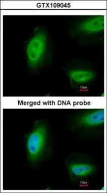
Immunofluorescence analysis of paraformaldehyde-fixed HeLa, using p73 (GTX109045) antibody at 1:100 dilution.
![Untreated (–) and treated (+) 293T whole cell extracts (30 μg) were separated by 7.5% SDS-PAGE, and the membrane was blotted with p73 antibody [N1C1] (GTX109045) diluted at 1:2000. The HRP-conjugated anti-rabbit IgG antibody (GTX213110-01) was used to detect the primary antibody, and the signal was developed with Trident ECL plus-Enhanced.](https://www.genetex.com/upload/website/prouct_img/normal/GTX109045/GTX109045_40044_20200131_WB_treatment_Doxorubicin_w_23060120_622.webp "Untreated (–) and treated (+) 293T whole cell extracts (30 μg) were separated by 7.5% SDS-PAGE, and the membrane was blotted with p73 antibody [N1C1] (GTX109045) diluted at 1:2000. The HRP-conjugated anti-rabbit IgG antibody (GTX213110-01) was used to detect the primary antibody, and the signal was developed with Trident ECL plus-Enhanced.")
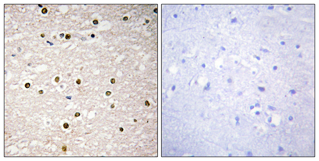
![p73 antibody [N1C1] detects p73 protein at nucleus in human colon carcinoma by immunohistochemical analysis. Sample: Paraffin-embedded human colon carcinoma. p73 antibody [N1C1] (GTX109045) diluted at 1:250.
Antigen Retrieval: Trilogy? (EDTA based, pH 8.0) buffer, 15min](https://www.genetex.com/upload/website/prouct_img/normal/GTX109045/GTX109045_40044_20151223_IHC-P_w_23060120_237.webp "p73 antibody [N1C1] detects p73 protein at nucleus in human colon carcinoma by immunohistochemical analysis. Sample: Paraffin-embedded human colon carcinoma. p73 antibody [N1C1] (GTX109045) diluted at 1:250.
Antigen Retrieval: Trilogy? (EDTA based, pH 8.0) buffer, 15min")
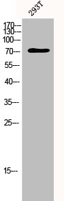
![Whole cell extract (30 μg) was separated by 7.5% SDS-PAGE, and the membrane was blotted with p73 antibody [N1C1] (GTX109045) diluted at 1:1000. The HRP-conjugated anti-rabbit IgG antibody (GTX213110-01) was used to detect the primary antibody.](https://www.genetex.com/upload/website/prouct_img/normal/GTX109045/GTX109045_40044_20200103_WB_w_23060120_284.webp "Whole cell extract (30 μg) was separated by 7.5% SDS-PAGE, and the membrane was blotted with p73 antibody [N1C1] (GTX109045) diluted at 1:1000. The HRP-conjugated anti-rabbit IgG antibody (GTX213110-01) was used to detect the primary antibody.")
 antibody at 1:100 dilution.
Antigen Retrieval: Trilogy? (EDTA based, pH 8.0) buffer, 15min")
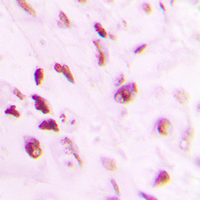
![p73 antibody [N1C1] detects p73 protein at nucleus in human SCC xenograft by immunohistochemical analysis. Sample: Paraffin-embedded human SCC xenograft . p73 antibody [N1C1] (GTX109045) diluted at 1:250.
Antigen Retrieval: Trilogy? (EDTA based, pH 8.0) buffer, 15min](https://www.genetex.com/upload/website/prouct_img/normal/GTX109045/GTX109045_40044_20151227_IHC-P_w_23060120_432.webp "p73 antibody [N1C1] detects p73 protein at nucleus in human SCC xenograft by immunohistochemical analysis. Sample: Paraffin-embedded human SCC xenograft . p73 antibody [N1C1] (GTX109045) diluted at 1:250.
Antigen Retrieval: Trilogy? (EDTA based, pH 8.0) buffer, 15min")
Immunofluorescence analysis of paraformaldehyde-fixed HeLa, using p73 (GTX109045) antibody at 1:100 dilution.
p73 antibody [N1C1]
GTX109045
ApplicationsImmunoFluorescence, Western Blot, ImmunoCytoChemistry, ImmunoHistoChemistry, ImmunoHistoChemistry Paraffin
Product group Antibodies
ReactivityHuman
TargetTP73
Overview
- SupplierGeneTex
- Product Namep73 antibody [N1C1]
- Delivery Days Customer9
- Application Supplier NoteWB: 1:500-1:3000. ICC/IF: 1:100-1:1000. IHC-P: 1:100-1:1000. *Optimal dilutions/concentrations should be determined by the researcher.Not tested in other applications.
- ApplicationsImmunoFluorescence, Western Blot, ImmunoCytoChemistry, ImmunoHistoChemistry, ImmunoHistoChemistry Paraffin
- CertificationResearch Use Only
- ClonalityPolyclonal
- Concentration0.89 mg/ml
- ConjugateUnconjugated
- Gene ID7161
- Target nameTP73
- Target descriptiontumor protein p73
- Target synonymsCILD47, P73, tumor protein p73, p53-like transcription factor, p53-related protein
- HostRabbit
- IsotypeIgG
- Protein IDO15350
- Protein NameTumor protein p73
- Scientific DescriptionThis gene encodes tumor protein p73, which is a member of the p53 family of transcription factors involved in cellular responses to stress and development. The family members include p53, p63, and p73 and have high sequence similarity to one another, which allows p63 and p73 to transactivate p53-responsive genes causing cell cycle arrest and apoptosis. The family members can interact with each other in many ways involving direct or indirect protein interactions, resulting in regulation of the same target gene promoters or regulation of each others promoters. The p73 protein is expressed at very low levels in normal tissues and is differentially expressed in a number of tumors. The p73 gene expresses at least 35 mRNA variants due to the use of alternate promoters, alternate translation initiation sites, and multiple splice variations. Theoretically this can account for 29 different p73 isoforms; however, the biological validity and the full-length nature of most variants have not been determined. [provided by RefSeq]
- ReactivityHuman
- Storage Instruction-20°C or -80°C,2°C to 8°C
- UNSPSC41116161
Datasheet
Related products
Product group Antibodies
Anti-p73 AntibodyA100047
ApplicationsWestern Blot, ELISA, ImmunoHistoChemistry
ReactivityHuman
- SizePrice
Product group Antibodies
Anti-TP73 Antibody144-61611
ApplicationsWestern Blot
ReactivityHuman, Mouse, Rat
TargetTP73
- SizePrice
Product group Antibodies

TP73 / p73 AntibodyLS-C831425
ApplicationsImmunoHistoChemistry
ReactivityHuman, Mouse
TargetTP73
- SizePrice
Product group Antibodies
P73 Recombinant Antibody, AbBy Fluor-647 ConjugatedBSM-62474R-BF647
ApplicationsImmunoFluorescence, Western Blot
ReactivityHuman, Mouse, Rat
TargetTP73
- SizePrice
Product group Antibodies
TP73 AntibodyCSB-PA003696
ApplicationsWestern Blot, ELISA
ReactivityHuman, Mouse
TargetTP73
- SizePrice
Product group Antibodies
TP73 Polyclonal AntibodyCAC15723
ApplicationsWestern Blot, ELISA, ImmunoHistoChemistry
TargetTP73
- SizePrice

Product group Antibodies
Anti-TP73 AntibodyHPA044516
ApplicationsWestern Blot, ChIP Chromatin ImmunoPrecipitation, ImmunoCytoChemistry
ReactivityHuman
TargetTP73
- SizePrice
![WB analysis of HeLa cell lysate using GTX46123 p73 antibody [5B429].](https://www.genetex.com/upload/website/prouct_img/normal/GTX46123/GTX46123_1642_WB_w_23060820_625.webp)
Product group Antibodies
p73 antibody [5B429]GTX46123
ApplicationsFlow Cytometry, ImmunoFluorescence, ImmunoPrecipitation, Mass Spectrometry, Western Blot, ChIP Chromatin ImmunoPrecipitation, ImmunoCytoChemistry, ImmunoHistoChemistry, ImmunoHistoChemistry Frozen, ImmunoHistoChemistry Paraffin
ReactivityHuman, Mouse, Rat
TargetTP73
- SizePrice
Product group Antibodies
p73 (phospho Tyr99) antibodyGTX50132
ApplicationsWestern Blot
ReactivityHuman
TargetTP73
- SizePrice
Product group Antibodies
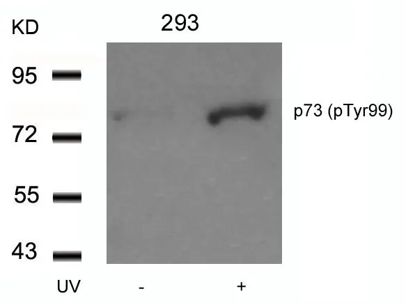
p73 (phospho Tyr99) antibodyGTX32277
ApplicationsWestern Blot, ImmunoHistoChemistry, ImmunoHistoChemistry Paraffin
ReactivityHuman
TargetTP73
- SizePrice