

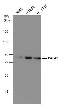
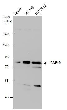
Various whole cell extracts (30 μg) were separated by 7.5% SDS-PAGE, and the membrane was blotted with PAF49 antibody (GTX629115) diluted at 1:500.
![PAF49 antibody [GT1964] detects PPY protein by Western blot analysis. A. 30 μg 293T whole cell lysate/extract B. 30 μg A431 whole cell lysate/extract C. 30 μg HeLa whole cell lysate/extract D. 30 μg HepG2 whole cell lysate/extract E. 30 μg A375 whole cell lysate/extract 7.5 % SDS-PAGE PAF49 antibody [GT1964] (GTX629115) dilution: 1:1000](https://www.genetex.com/upload/website/prouct_img/normal/GTX629115/GTX629115_41309_WB_w_23061202_582.webp "PAF49 antibody [GT1964] detects PPY protein by Western blot analysis. A. 30 μg 293T whole cell lysate/extract B. 30 μg A431 whole cell lysate/extract C. 30 μg HeLa whole cell lysate/extract D. 30 μg HepG2 whole cell lysate/extract E. 30 μg A375 whole cell lysate/extract 7.5 % SDS-PAGE PAF49 antibody [GT1964] (GTX629115) dilution: 1:1000")
 diluted at 1:500. Blue: Hoechst 33342 staining.")
Various whole cell extracts (30 μg) were separated by 7.5% SDS-PAGE, and the membrane was blotted with PAF49 antibody (GTX629115) diluted at 1:500.
PAF49 antibody [GT1964]
GTX629115
ApplicationsImmunoFluorescence, Western Blot, ImmunoCytoChemistry
Product group Antibodies
ReactivityHuman
TargetPOLR1G
Overview
- SupplierGeneTex
- Product NamePAF49 antibody [GT1964]
- Delivery Days Customer9
- Application Supplier NoteWB: 1:500-1:3000. ICC/IF: 1:100-1:1000. *Optimal dilutions/concentrations should be determined by the researcher.Not tested in other applications.
- ApplicationsImmunoFluorescence, Western Blot, ImmunoCytoChemistry
- CertificationResearch Use Only
- ClonalityMonoclonal
- Clone IDGT1964
- Concentration1 mg/ml
- ConjugateUnconjugated
- Gene ID10849
- Target namePOLR1G
- Target descriptionRNA polymerase I subunit G
- Target synonymsASE-1, ASE1, CAST, CD3EAP, PAF49, RPA34, DNA-directed RNA polymerase I subunit RPA34, CD3e antigen, epsilon polypeptide associated protein, CD3e molecule associated protein, CD3e molecule, epsilon associated protein, DNA-directed RNA polymerase I subunit G, RNA polymerase I-associated factor PAF49, antisense to ERCC-1 protein
- HostMouse
- IsotypeIgG2b
- Protein IDO15446
- Protein NameDNA-directed RNA polymerase I subunit RPA34
- ReactivityHuman
- Storage Instruction-20°C or -80°C,2°C to 8°C
- UNSPSC41116161
Datasheet
Related products
Product group Antibodies
CD3EAP AntibodyCSB-PA004932LA01HU
ApplicationsImmunoFluorescence, Western Blot, ELISA, ImmunoHistoChemistry
ReactivityHuman, Rat
TargetPOLR1G
- SizePrice
Product group Antibodies
Anti-CD3EAP AntibodyA100594
ApplicationsWestern Blot, ELISA
ReactivityHuman
- SizePrice

Product group Antibodies
ApplicationsImmunoFluorescence, Western Blot, ELISA, ImmunoCytoChemistry, ImmunoHistoChemistry
TargetPOLR1G
- SizePrice

Product group Antibodies
Anti-CD3EAP AntibodyHPA041664
ApplicationsWestern Blot, ImmunoCytoChemistry, ImmunoHistoChemistry
ReactivityHuman
TargetPOLR1G
- SizePrice
Product group Antibodies

Goat anti-CD3EAPEB12596
ApplicationsELISA
ReactivityHuman
TargetPOLR1G
- SizePrice
Product group Antibodies

CD3EAP AntibodyLS-C672546
ApplicationsImmunoFluorescence, Western Blot, ELISA, ImmunoHistoChemistry, ImmunoHistoChemistry Paraffin
ReactivityHuman
TargetPOLR1G
- SizePrice
Product group Antibodies
CD3EAP Polyclonal AntibodyCAC15092
ApplicationsImmunoFluorescence, Western Blot, ELISA, ImmunoHistoChemistry
ReactivityRat
TargetPOLR1G
- SizePrice
![PAF49 antibody detects PAF49 protein at nucleolus by immunofluorescent analysis. Sample: HeLa cells were fixed in ice-cold MeOH for 5 min. Green: PAF49 protein stained by PAF49 antibody (GTX102175) diluted at 1:1000. Red: alpha Tubulin, a cytoskeleton marker, stained by alpha Tubulin antibody [GT114] (GTX628802) diluted at 1:500. Blue: Hoechst 33342 staining.](https://www.genetex.com/upload/website/prouct_img/normal/GTX102175/GTX102175_42634_20161228_IFA_w_23060100_721.webp)
Product group Antibodies
PAF49 antibodyGTX102175
ApplicationsImmunoFluorescence, Western Blot, ImmunoCytoChemistry, ImmunoHistoChemistry, ImmunoHistoChemistry Paraffin
ReactivityHuman
TargetPOLR1G
- SizePrice
Product group Antibodies
PAF49 antibody [GT7212]GTX629075
ApplicationsImmunoFluorescence, Western Blot, ImmunoCytoChemistry
ReactivityHuman
TargetPOLR1G
- SizePrice
![Non-transfected (–) and transfected (+) 293T whole cell extracts (50 μg) were separated by 7.5% SDS-PAGE, and the membrane was blotted with PAF49 antibody [GT635] (GTX629076) diluted at 1:1000. The HRP-conjugated anti-mouset IgG antibody (GTX213111-01) was used to detect the primary antibody.](https://www.genetex.com/upload/website/prouct_img/normal/GTX629076/GTX629076_41309_20170511_WB_shRNA_watermark_w_23061202_478.webp)
Product group Antibodies
PAF49 antibody [GT635]GTX629076
ApplicationsImmunoFluorescence, Western Blot, ImmunoCytoChemistry
ReactivityHuman
TargetPOLR1G
- SizePrice