

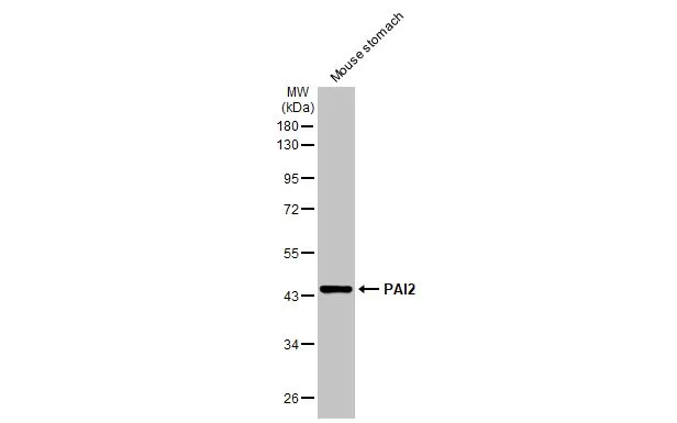
Mouse tissue extract (50 μg) was separated by 10% SDS-PAGE, and the membrane was blotted with PAI2 antibody (GTX103194) diluted at 1:1000. The HRP-conjugated anti-rabbit IgG antibody (GTX213110-01) was used to detect the primary antibody, and the signal was developed with Trident ECL plus-Enhanced.
 antibody at 1:500 dilution.
Antigen Retrieval: Trilogy? (EDTA based, pH 8.0) buffer, 15min")
 diluted at 1:500. Blue: Hoechst 33342 staining. Scale bar = 10 μm.")
 A: U87-MG 10% SDS PAGE GTX103194 diluted at 1:10000 The HRP-conjugated anti-rabbit IgG antibody (GTX213110-01) was used to detect the primary antibody.")
 were separated by 10% SDS-PAGE, and the membrane was blotted with PAI2 antibody (GTX103194) diluted at a dilution of 1:1000. The HRP-conjugated anti-rabbit IgG antibody (GTX213110-01) was used to detect the primary antibody.")
. Western blot analysis was performed using PAI2 antibody (GTX103194). EasyBlot anti-Rabbit IgG (GTX221666-01) was used as a secondary reagent.")
Mouse tissue extract (50 μg) was separated by 10% SDS-PAGE, and the membrane was blotted with PAI2 antibody (GTX103194) diluted at 1:1000. The HRP-conjugated anti-rabbit IgG antibody (GTX213110-01) was used to detect the primary antibody, and the signal was developed with Trident ECL plus-Enhanced.
PAI2 antibody
GTX103194
ApplicationsImmunoFluorescence, ImmunoPrecipitation, Western Blot, ImmunoCytoChemistry, ImmunoHistoChemistry, ImmunoHistoChemistry Paraffin
Product group Antibodies
ReactivityHuman, Mouse
TargetSERPINB2
Overview
- SupplierGeneTex
- Product NamePAI2 antibody
- Delivery Days Customer9
- Application Supplier NoteWB: 1:500-1:20000. ICC/IF: 1:100-1:1000. IHC-P: 1:100-1:1000. IP: 1:100-1:500. *Optimal dilutions/concentrations should be determined by the researcher.Not tested in other applications.
- ApplicationsImmunoFluorescence, ImmunoPrecipitation, Western Blot, ImmunoCytoChemistry, ImmunoHistoChemistry, ImmunoHistoChemistry Paraffin
- CertificationResearch Use Only
- ClonalityPolyclonal
- Concentration0.39 mg/ml
- ConjugateUnconjugated
- Gene ID5055
- Target nameSERPINB2
- Target descriptionserpin family B member 2
- Target synonymsHsT1201, PAI, PAI-2, PAI2, PLANH2, plasminogen activator inhibitor 2, monocyte Arg-serpin, placental plasminogen activator inhibitor, plasminogen activator inhibitor, type II (arginine-serpin), plasminogen-activator inhibitor, serine (or cysteine) proteinase inhibitor, clade B (ovalbumin), member 2, serpin B2, serpin peptidase inhibitor, clade B (ovalbumin), member 2, urokinase inhibitor
- HostRabbit
- IsotypeIgG
- Protein IDP05120
- Protein NamePlasminogen activator inhibitor 2
- Scientific DescriptionInhibits urokinase-type plasminogen activator. The monocyte derived PAI-2 is distinct from the endothelial cell-derived PAI-1.
- ReactivityHuman, Mouse
- Storage Instruction-20°C or -80°C,2°C to 8°C
- UNSPSC41116161
Datasheet
Related products
Product group Antibodies
Anti-SERPINB2 AntibodyA44998
ApplicationsImmunoHistoChemistry
ReactivityHuman
- SizePrice
Product group Antibodies
Anti-SERPINB2 Antibody144-60984
ApplicationsWestern Blot
ReactivityHuman
TargetSERPINB2
- SizePrice
Product group Antibodies

SERPINB2 / PAI-2 AntibodyLS-C831580
ApplicationsImmunoHistoChemistry
ReactivityHuman, Mouse, Rat
TargetSERPINB2
- SizePrice
Product group Antibodies
SerpinB2 Polyclonal AntibodyBS-1864R
ApplicationsImmunoFluorescence, ELISA, ImmunoCytoChemistry, ImmunoHistoChemistry, ImmunoHistoChemistry Frozen, ImmunoHistoChemistry Paraffin
ReactivityBovine, Canine, Human, Mouse, Porcine, Rabbit, Rat
TargetSERPINB2
- SizePrice
Product group Antibodies
SERPINB2 Polyclonal AntibodyCAC15655
ApplicationsWestern Blot, ELISA, ImmunoHistoChemistry
TargetSERPINB2
- SizePrice
Product group Antibodies
SERPINB2 AntibodyCSB-PA020201
ApplicationsWestern Blot, ELISA, ImmunoHistoChemistry
ReactivityHuman
TargetSERPINB2
- SizePrice
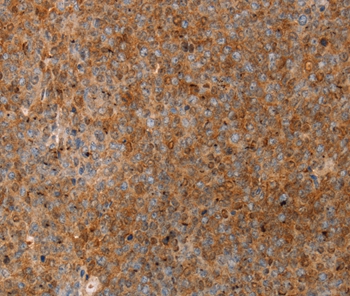
![PAI2 antibody [HL2218] detects secreted PAI2 protein by immunohistochemical analysis. Sample: Paraffin-embedded mouse stomach. PAI2 stained by PAI2 antibody [HL2218] (GTX638222) diluted at 1:100. Antigen Retrieval: Citrate buffer, pH 6.0, 15 min](https://www.genetex.com/upload/website/prouct_img/normal/GTX638222/GTX638222_T-44942_20230303_IHC-P_M_23031402_331.webp)
Product group Antibodies
PAI2 antibody [HL2218]GTX638222
ApplicationsWestern Blot, ImmunoHistoChemistry, ImmunoHistoChemistry Paraffin
ReactivityHuman, Mouse
TargetSERPINB2
- SizePrice
![IHC-P analysis of human placenta (early) tissue using GTX640562 PAI2 antibody [HMV330] HistoMAX?. Strong cytoplasmic and nuclear PAI2 staining of trophoblast cells.](https://www.genetex.com/upload/website/prouct_img/normal/GTX640562/GTX640562_20240703_IHC-P_24070300_228.webp)
Product group Antibodies
PAI2 antibody [HMV330] HistoMAX(tm)GTX640562
ApplicationsImmunoHistoChemistry, ImmunoHistoChemistry Paraffin
ReactivityHuman
TargetSERPINB2
- SizePrice

Product group Antibodies
Anti-SERPINB2 AntibodyHPA015480
ApplicationsImmunoHistoChemistry
ReactivityHuman
TargetSERPINB2
- SizePrice

Product group Antibodies
Anti-SerpinB2 Antibody Picoband(r)PB9355-CARRIER-FREE
ApplicationsWestern Blot, ImmunoHistoChemistry
ReactivityHuman
TargetSERPINB2
- SizePrice