

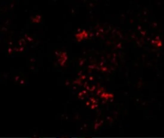
IHC-P analysis of mouse spleen tissue using GTX31742 PAK2 antibody. Working concentration : 20 μg/ml

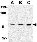
 0.5 and (B) 1 μg/ml")
IHC-P analysis of mouse spleen tissue using GTX31742 PAK2 antibody. Working concentration : 20 μg/ml
PAK2 antibody
GTX31742
ApplicationsWestern Blot, ELISA, ImmunoHistoChemistry, ImmunoHistoChemistry Paraffin
Product group Antibodies
ReactivityHuman, Mouse, Rat
TargetPAK2
Overview
- SupplierGeneTex
- Product NamePAK2 antibody
- Delivery Days Customer9
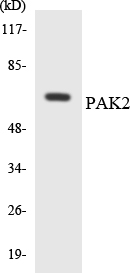
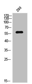
- Application Supplier NoteWB: 0.5 - 1 microg/mL. IHC-P: 10 microg/mL. *Optimal dilutions/concentrations should be determined by the researcher.Not tested in other applications.
- ApplicationsWestern Blot, ELISA, ImmunoHistoChemistry, ImmunoHistoChemistry Paraffin
- CertificationResearch Use Only
- ClonalityPolyclonal
- Concentration1 mg/ml
- ConjugateUnconjugated
- Gene ID5062
- Target namePAK2
- Target descriptionp21 (RAC1) activated kinase 2
- Target synonymsKNO2, PAK65, PAKgamma, serine/threonine-protein kinase PAK 2, PAK-2, S6/H4 kinase, gamma-PAK, p21 (CDKN1A)-activated kinase 2, p21 protein (Cdc42/Rac)-activated kinase 2, p21-activated kinase 2, p58
- HostRabbit
- IsotypeIgG
- Protein IDQ13177
- Protein NameSerine/threonine-protein kinase PAK 2
- Scientific DescriptionThe p21 activated kinases (PAK) are critical effectors that link Rho GTPases to cytoskeleton reorganization and nuclear signaling. The PAK proteins are a family of serine/threonine kinases that serve as targets for the small GTP binding proteins, CDC42 and RAC1, and have been implicated in a wide range of biological activities. The protein encoded by this gene is activated by proteolytic cleavage during caspase-mediated apoptosis, and may play a role in regulating the apoptotic events in the dying cell. [provided by RefSeq, Jul 2008]
- ReactivityHuman, Mouse, Rat
- Storage Instruction-20°C or -80°C,2°C to 8°C
- UNSPSC41116161
Datasheet
Related products
Product group Antibodies
Anti-PAK2 AntibodyA96147
ApplicationsWestern Blot, ELISA, ImmunoHistoChemistry
ReactivityHuman, Mouse, Rat
- SizePrice
Product group Antibodies
Anti-PAK2 Antibody144-07333
ApplicationsImmunoFluorescence, Western Blot, ImmunoHistoChemistry
ReactivityHuman, Mouse, Rat
TargetPAK2
- SizePrice

Product group Antibodies
Anti-PAK2 Antibody Picoband(r)A01419-4-CARRIER-FREE
ApplicationsFlow Cytometry, ImmunoFluorescence, Western Blot, ELISA, ImmunoCytoChemistry, ImmunoHistoChemistry
ReactivityHuman, Mouse, Rat
TargetPAK2
- SizePrice
Product group Antibodies
PAK2 Recombinant Antibody, Biotin ConjugatedBSM-61673R-BIOTIN
ApplicationsWestern Blot, ImmunoHistoChemistry, ImmunoHistoChemistry Frozen, ImmunoHistoChemistry Paraffin
ReactivityHuman, Mouse, Rat
TargetPAK2
- SizePrice
Product group Antibodies
Phospho-PAK2 (S20) AntibodyCSB-PA010536
ApplicationsWestern Blot, ELISA, ImmunoHistoChemistry
ReactivityHuman, Mouse, Rat
TargetPAK2
- SizePrice
Product group Antibodies
ApplicationsWestern Blot, ImmunoHistoChemistry
ReactivityMouse
TargetPAK2
- SizePrice
Product group Antibodies

PAK2 AntibodyLS-C400945
ApplicationsELISA, ImmunoHistoChemistry
ReactivityHuman, Mouse, Rat
TargetPAK2
- SizePrice
Product group Antibodies
PAK2 antibodyGTX31743
ApplicationsWestern Blot, ELISA, ImmunoHistoChemistry, ImmunoHistoChemistry Paraffin
ReactivityHuman, Mouse, Rat
TargetPAK2
- SizePrice
![ICC/IF analysis of HeLa cells using GTX83373 PAK2 antibody [3B5]. Green : PAK2 Blue: DRAQ5 fluorescent DNA dye](https://www.genetex.com/upload/website/prouct_img/normal/GTX83373/GTX83373_20170912_ICCIF_w_23061322_871.webp)
Product group Antibodies
PAK2 antibody [3B5]GTX83373
ApplicationsImmunoFluorescence, Western Blot, ELISA, ImmunoCytoChemistry, ImmunoHistoChemistry, ImmunoHistoChemistry Paraffin
ReactivityHuman, Monkey
TargetPAK2
- SizePrice
Product group Antibodies
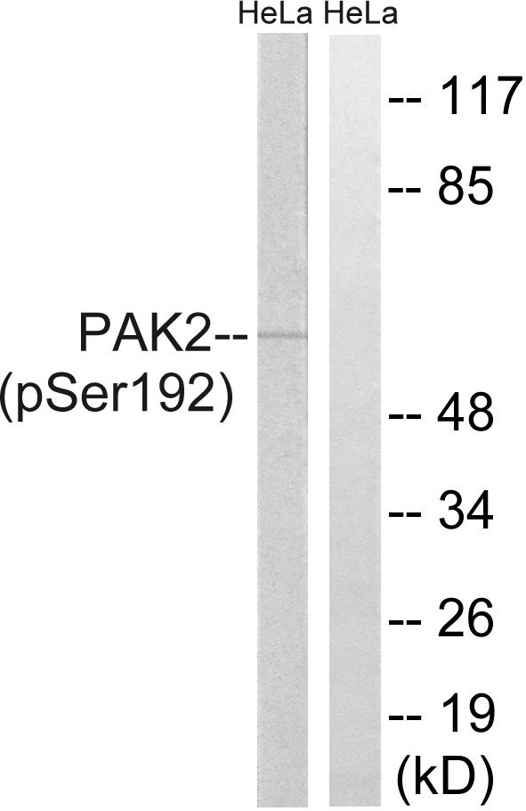
PAK2 (phospho Ser192) antibodyGTX86345
ApplicationsWestern Blot
ReactivityHuman
TargetPAK2
- SizePrice