

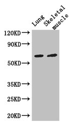
Western Blot Positive WB detected in: Mouse lung tissue, Mouse skeletal muscle tissue All lanes: PAK3 antibody at 3ug/ml Secondary Goat polyclonal to rabbit IgG at 1/50000 dilution Predicted band size: 63, 61, 65 kDa Observed band size: 63 kDa

")
Western Blot Positive WB detected in: Mouse lung tissue, Mouse skeletal muscle tissue All lanes: PAK3 antibody at 3ug/ml Secondary Goat polyclonal to rabbit IgG at 1/50000 dilution Predicted band size: 63, 61, 65 kDa Observed band size: 63 kDa
PAK3 Antibody
CSB-PA017407LA01HU
ApplicationsImmunoFluorescence, Western Blot, ELISA, ImmunoHistoChemistry
Product group Antibodies
ReactivityHuman, Mouse
TargetPAK3
Overview
- SupplierCusabio
- Product NamePAK3 Antibody
- Delivery Days Customer20
- ApplicationsImmunoFluorescence, Western Blot, ELISA, ImmunoHistoChemistry
- CertificationResearch Use Only
- ClonalityPolyclonal
- ConjugateUnconjugated
- Gene ID5063
- Target namePAK3
- Target descriptionp21 (RAC1) activated kinase 3
- Target synonymsARA, MRX30, MRX47, OPHN3, PAK-3, PAK3beta, XLID30, bPAK, beta-PAK, serine/threonine-protein kinase PAK 3, adriamycin resistance-associated, oligophrenin-3, p21 (CDKN1A)-activated kinase 3, p21 protein (Cdc42/Rac)-activated kinase 3
- HostRabbit
- IsotypeIgG
- Protein IDO75914
- Protein NameSerine/threonine-protein kinase PAK 3
- Scientific DescriptionSerine/threonine protein kinase that plays a role in a variety of different signaling pathways including cytoskeleton regulation, cell migration, or cell cycle regulation. Plays a role in dendrite spine morphogenesis as well as synapse formation and plasticity. Acts as downstream effector of the small GTPases CDC42 and RAC1. Activation by the binding of active CDC42 and RAC1 results in a conformational change and a subsequent autophosphorylation on several serine and/or threonine residues. Phosphorylates MAPK4 and MAPK6 and activates the downstream target MAPKAPK5, a regulator of F-actin polymerization and cell migration. Additionally, phosphorylates TNNI3/troponin I to modulate calcium sensitivity and relaxation kinetics of thin myofilaments. May also be involved in early neuronal development.
- ReactivityHuman, Mouse
- Storage Instruction-20°C or -80°C
- UNSPSC41116161
Related products
Product group Antibodies
Anti-PAK3 AntibodyA97982
ApplicationsWestern Blot, ELISA
ReactivityHuman, Mouse, Rat
- SizePrice

Product group Antibodies
Anti-PAK3 Antibody Picoband(r)A03124-1-CARRIER-FREE
ApplicationsFlow Cytometry, ImmunoFluorescence, Western Blot, ELISA, ImmunoCytoChemistry, ImmunoHistoChemistry
ReactivityHuman, Mouse, Rat
TargetPAK3
- SizePrice
Product group Antibodies
Anti-PAK3 Antibody144-64728
ApplicationsImmunoFluorescence, Western Blot
ReactivityHuman, Mouse, Rat
TargetPAK3
- SizePrice
Product group Antibodies
PAK3 Recombinant Antibody, AbBy Fluor-350 ConjugatedBSM-61672R-BF350
ApplicationsImmunoFluorescence, Western Blot, ImmunoCytoChemistry
ReactivityHuman, Mouse, Rat
TargetPAK3
- SizePrice
Product group Antibodies
PAK3 Polyclonal AntibodyCAC13147
ApplicationsImmunoFluorescence, Western Blot, ELISA, ImmunoHistoChemistry
ReactivityMouse
TargetPAK3
- SizePrice
Product group Antibodies

PAK3 Antibody (Internal)LS-C368453
ApplicationsWestern Blot, ImmunoHistoChemistry, ImmunoHistoChemistry Paraffin
ReactivityHuman, Monkey, Mouse, Rat
TargetPAK3
- SizePrice

Product group Antibodies
Anti-PAK3 AntibodyHPA060219
ApplicationsWestern Blot, ImmunoHistoChemistry
ReactivityHuman
TargetPAK3
- SizePrice
Product group Antibodies
PAK3 antibodyGTX109991
ApplicationsWestern Blot
ReactivityHuman, Mouse, Rat
TargetPAK3
- SizePrice
Product group Antibodies
ApplicationsWestern Blot, ELISA, ImmunoHistoChemistry, ImmunoHistoChemistry Paraffin
ReactivityHuman
TargetPAK3
- SizePrice