

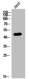
PAX1 Antibody (aa216-265, HRP)
LS-C464505
ApplicationsWestern Blot
Product group Antibodies
TargetPAX1
Overview
- SupplierLifeSpan BioSciences
- Product NamePAX1 Antibody (aa216-265, HRP)
- Delivery Days Customer23
- Application Supplier NoteThe applications listed have been tested for the unconjugated form of this product. Other forms have not been tested.
- ApplicationsWestern Blot
- Applications SupplierWB The applications listed have been tested for the unconjugated form of this product. Other forms have not been tested.
- CertificationResearch Use Only
- ClonalityPolyclonal
- Concentration0.65 mg/ml
- ConjugateHRP
- Estimated Purity...
- Gene ID5075
- Target namePAX1
- Target descriptionpaired box 1
- Target synonymsHUP48; OFC2; paired box gene 1; paired box protein Pax-1; paired domain gene HuP48
- HostRabbit
- IsotypeIgG
- Storage Instruction-20°C,2°C to 8°C
- UNSPSC12352203
Related products
Product group Antibodies
PAX1 AntibodyCSB-PA020238
ApplicationsWestern Blot, ELISA, ImmunoHistoChemistry
ReactivityHuman
TargetPAX1
- SizePrice


Product group Antibodies
Anti-PAX1 Antibody Picoband(r)A04559-2-CARRIER-FREE
ApplicationsFlow Cytometry, Western Blot, ELISA
TargetPAX1
- SizePrice
Product group Antibodies

Goat anti-PAX1 (aa318-329)EB10702
ApplicationsWestern Blot, ELISA
TargetPAX1
- SizePrice
Product group Antibodies
PAX1 antibodyGTX02870
ApplicationsWestern Blot, ImmunoHistoChemistry, ImmunoHistoChemistry Paraffin
TargetPAX1
- SizePrice
Product group Antibodies
PAX1 Polyclonal AntibodyBS-1199R
ApplicationsELISA, ImmunoHistoChemistry, ImmunoHistoChemistry Frozen, ImmunoHistoChemistry Paraffin
TargetPAX1
- SizePrice

Product group Antibodies
Anti-Pax-1 AntibodyA99612
ApplicationsWestern Blot, ELISA, ImmunoHistoChemistry
- SizePrice