

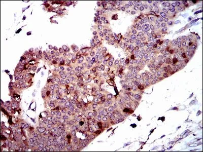
IHC-P analysis of ovarian cancer tissue using GTX60560 PBK antibody [2C8].
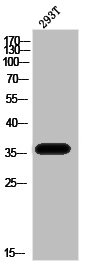
![WB analysis of A431 cell lysate using GTX60560 PBK antibody [2C8].](https://www.genetex.com/upload/website/prouct_img/normal/GTX60560/GTX60560_20170912_WB_w_23061123_267.webp "WB analysis of A431 cell lysate using GTX60560 PBK antibody [2C8].")
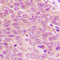
![IHC-P analysis of colon cancer tissue using GTX60560 PBK antibody [2C8].](https://www.genetex.com/upload/website/prouct_img/normal/GTX60560/GTX60560_20170912_IHC-P_w_23061123_951.webp "IHC-P analysis of colon cancer tissue using GTX60560 PBK antibody [2C8].")
![WB analysis of human PBK (AA: 50-230) recombinant protein using GTX60560 PBK antibody [2C8].](https://www.genetex.com/upload/website/prouct_img/normal/GTX60560/GTX60560_20170912_WB_1_w_23061123_117.webp "WB analysis of human PBK (AA: 50-230) recombinant protein using GTX60560 PBK antibody [2C8].")
![ELISA analysis of antigen using GTX60560 PBK antibody [2C8].
Black : Control antigen 100ng
Purple : Antigen 10ng
Blue : Antigen 50ng
Red : Antigen 100ng](https://www.genetex.com/upload/website/prouct_img/normal/GTX60560/GTX60560_20170912_ELISA_w_23061123_612.webp "ELISA analysis of antigen using GTX60560 PBK antibody [2C8].
Black : Control antigen 100ng
Purple : Antigen 10ng
Blue : Antigen 50ng
Red : Antigen 100ng")
![FACS analysis of HeLa cells using GTX60560 PBK antibody [2C8]. Green : PBK Red : negative control](https://www.genetex.com/upload/website/prouct_img/normal/GTX60560/GTX60560_20170912_FACS_w_23061123_344.webp "FACS analysis of HeLa cells using GTX60560 PBK antibody [2C8]. Green : PBK Red : negative control")
![ICC/IF analysis of HeLa cells using GTX60560 PBK antibody [2C8]. Green : PBK Blue: DRAQ5 fluorescent DNA dye Red: Actin filaments](https://www.genetex.com/upload/website/prouct_img/normal/GTX60560/GTX60560_20170912_ICCIF_w_23061123_599.webp "ICC/IF analysis of HeLa cells using GTX60560 PBK antibody [2C8]. Green : PBK Blue: DRAQ5 fluorescent DNA dye Red: Actin filaments")
IHC-P analysis of ovarian cancer tissue using GTX60560 PBK antibody [2C8].
PBK antibody [2C8]
GTX60560
ApplicationsFlow Cytometry, ImmunoFluorescence, Western Blot, ELISA, ImmunoCytoChemistry, ImmunoHistoChemistry, ImmunoHistoChemistry Paraffin
Product group Antibodies
ReactivityHuman
TargetPBK
Overview
- SupplierGeneTex
- Product NamePBK antibody [2C8]
- Delivery Days Customer9
- Application Supplier NoteWB: 1/500 - 1/2000. ICC/IF: 1/200 - 1/1000. IHC-P: 1/200 - 1/1000. FCM: 1/200 - 1/400. ELISA: 1/10000. *Optimal dilutions/concentrations should be determined by the researcher.Not tested in other applications.
- ApplicationsFlow Cytometry, ImmunoFluorescence, Western Blot, ELISA, ImmunoCytoChemistry, ImmunoHistoChemistry, ImmunoHistoChemistry Paraffin
- CertificationResearch Use Only
- ClonalityMonoclonal
- Clone ID2C8
- ConjugateUnconjugated
- Gene ID55872
- Target namePBK
- Target descriptionPDZ binding kinase
- Target synonymsCT84, HEL164, Nori-3, SPK, TOPK, lymphokine-activated killer T-cell-originated protein kinase, MAPKK-like protein kinase, T-LAK cell-originated protein kinase, cancer/testis antigen 84, epididymis luminal protein 164, serine/threonine protein kinase, spermatogenesis-related protein kinase
- HostMouse
- IsotypeIgG2b
- Protein IDQ96KB5
- Protein NameLymphokine-activated killer T-cell-originated protein kinase
- Scientific DescriptionThis genes encodes a serine/threonine kinase related to the dual specific mitogen-activated protein kinase kinase (MAPKK) family. Evidence suggests that mitotic phosphorylation is required for its catalytic activity. This mitotic kinase may be involved in the activation of lymphoid cells and support testicular functions, with a suggested role in the process of spermatogenesis. [provided by RefSeq, Jul 2008]
- ReactivityHuman
- Storage Instruction-20°C or -80°C,2°C to 8°C
- UNSPSC41116161
References
- Targeting of RRM2 suppresses DNA damage response and activates apoptosis in atypical teratoid rhabdoid tumor.Read this paper
Datasheet
Related products
Product group Antibodies
Anti-PBK AntibodyA100038
ApplicationsWestern Blot, ELISA, ImmunoHistoChemistry
ReactivityHuman
- SizePrice
Product group Antibodies

SPK AntibodyABX123162
ApplicationsWestern Blot, ImmunoHistoChemistry
- SizePrice
Product group Antibodies

Anti-PBK [RAB-C423]Ab01833-1.1
ApplicationsImmunoPrecipitation
ReactivityHuman
TargetPBK
- SizePrice
Product group Antibodies

ApplicationsELISA
ReactivityHuman
TargetPBK
- SizePrice

Product group Antibodies
Anti-PBK AntibodyHPA005753
ApplicationsWestern Blot, ImmunoCytoChemistry, ImmunoHistoChemistry
ReactivityHuman, Mouse
TargetPBK
- SizePrice
Product group Antibodies
Phospho-PBK (T9) AntibodyCSB-PA030267
ApplicationsWestern Blot, ELISA, ImmunoHistoChemistry
ReactivityHuman
TargetPBK
- SizePrice

Product group Antibodies
Anti-PBK Antibody Picoband(r)PB9310-CARRIER-FREE
ApplicationsFlow Cytometry, ImmunoFluorescence, Western Blot, ImmunoCytoChemistry, ImmunoHistoChemistry
ReactivityHuman, Mouse, Rat
TargetPBK
- SizePrice
![Immunoprecipitation of PBK protein from A431 whole cell extracts using 5 μg of PBK antibody [N2C3] (GTX113982). Western blot analysis was performed using PBK antibody [N2C3] (GTX113982). EasyBlot anti-Rabbit IgG (GTX221666-01) was used as a secondary reagent.](https://www.genetex.com/upload/website/prouct_img/normal/GTX113982/GTX113982_40150_20151127_IP_w_23060501_943.webp)
Product group Antibodies
PBK antibody [N2C3]GTX113982
ApplicationsImmunoPrecipitation, Western Blot
ReactivityHuman
TargetPBK
- SizePrice
Product group Antibodies
PBK (phospho Thr9) antibodyGTX54960
ApplicationsWestern Blot, ImmunoHistoChemistry, ImmunoHistoChemistry Paraffin
ReactivityHuman
TargetPBK
- SizePrice
Product group Antibodies
PBK antibodyGTX33388
ApplicationsWestern Blot, ImmunoHistoChemistry, ImmunoHistoChemistry Paraffin
ReactivityHuman, Rat
TargetPBK
- SizePrice