

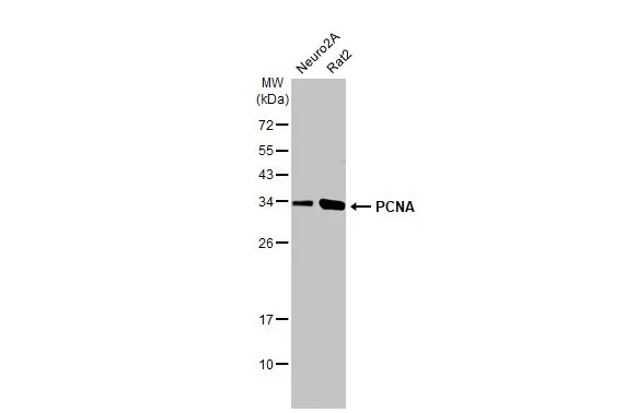
Various whole cell extracts (30 μg) were separated by 12% SDS-PAGE, and the membrane was blotted with PCNA antibody [HL1965] (GTX637858) diluted at 1:1000. The HRP-conjugated anti-rabbit IgG antibody (GTX213110-01) was used to detect the primary antibody.
![Various whole cell extracts (30 μg) were separated by 12% SDS-PAGE, and the membrane was blotted with PCNA antibody [HL1965] (GTX637858) diluted at 1:1000. The HRP-conjugated anti-rabbit IgG antibody (GTX213110-01) was used to detect the primary antibody.](https://www.genetex.com/upload/website/prouct_img/normal/GTX637858/GTX637858_T-44858_20221209_WB_D_C_22121123_903.webp "Various whole cell extracts (30 μg) were separated by 12% SDS-PAGE, and the membrane was blotted with PCNA antibody [HL1965] (GTX637858) diluted at 1:1000. The HRP-conjugated anti-rabbit IgG antibody (GTX213110-01) was used to detect the primary antibody.")
![Whole zebrafish extract (30 μg) was separated by 12% SDS-PAGE, and the membrane was blotted with PCNA antibody [HL1965] (GTX637858) diluted at 1:1000. The HRP-conjugated anti-rabbit IgG antibody (GTX213110-01) was used to detect the primary antibody.](https://www.genetex.com/upload/website/prouct_img/normal/GTX637858/GTX637858_T-44858_20221209_WB_Z_22121123_114.webp "Whole zebrafish extract (30 μg) was separated by 12% SDS-PAGE, and the membrane was blotted with PCNA antibody [HL1965] (GTX637858) diluted at 1:1000. The HRP-conjugated anti-rabbit IgG antibody (GTX213110-01) was used to detect the primary antibody.")
![Various whole cell extracts (30 μg) were separated by 12% SDS-PAGE, and the membrane was blotted with PCNA antibody [HL1965] (GTX637858) diluted at 1:1000. The HRP-conjugated anti-rabbit IgG antibody (GTX213110-01) was used to detect the primary antibody. Corresponding RNA expression data for the same cell lines are based on Human Protein Atlas program.](https://www.genetex.com/upload/website/prouct_img/normal/GTX637858/GTX637858_44907_20221230_WB_23010400_849.webp "Various whole cell extracts (30 μg) were separated by 12% SDS-PAGE, and the membrane was blotted with PCNA antibody [HL1965] (GTX637858) diluted at 1:1000. The HRP-conjugated anti-rabbit IgG antibody (GTX213110-01) was used to detect the primary antibody. Corresponding RNA expression data for the same cell lines are based on Human Protein Atlas program.")
![Various whole cell extracts (30 μg) were separated by 12% SDS-PAGE, and the membrane was blotted with PCNA antibody [HL1965] (GTX637858) diluted at 1:1000. The HRP-conjugated anti-rabbit IgG antibody (GTX213110-01) was used to detect the primary antibody.](https://www.genetex.com/upload/website/prouct_img/normal/GTX637858/GTX637858_44907_20240920_WB_24092600_190.webp "Various whole cell extracts (30 μg) were separated by 12% SDS-PAGE, and the membrane was blotted with PCNA antibody [HL1965] (GTX637858) diluted at 1:1000. The HRP-conjugated anti-rabbit IgG antibody (GTX213110-01) was used to detect the primary antibody.")
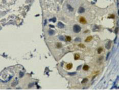
![PCNA antibody [HL1965] detects PCNA protein by immunohistochemical analysis. Sample: Frozen-sectioned mouse testis. Green: PCNA stained by PCNA antibody [HL1965] (GTX637858) diluted at 1:200. Red: Vimentin antibody [GT812] (GTX629744) diluted at 1:1000. Blue: Hoechst 33342 staining.](https://www.genetex.com/upload/website/prouct_img/normal/GTX637858/GTX637858_44907_20250704_IHC-Fr_M_25071023_502.webp "PCNA antibody [HL1965] detects PCNA protein by immunohistochemical analysis. Sample: Frozen-sectioned mouse testis. Green: PCNA stained by PCNA antibody [HL1965] (GTX637858) diluted at 1:200. Red: Vimentin antibody [GT812] (GTX629744) diluted at 1:1000. Blue: Hoechst 33342 staining.")
![Whole Japanese medaka extract (30 μg) was separated by 12% SDS-PAGE, and the membrane was blotted with PCNA antibody [HL1965] (GTX637858) diluted at 1:1000. The HRP-conjugated anti-rabbit IgG antibody (GTX213110-01) was used to detect the primary antibody.](https://www.genetex.com/upload/website/prouct_img/normal/GTX637858/GTX637858_44907_20250815_WB_medaka_25082121_368.webp "Whole Japanese medaka extract (30 μg) was separated by 12% SDS-PAGE, and the membrane was blotted with PCNA antibody [HL1965] (GTX637858) diluted at 1:1000. The HRP-conjugated anti-rabbit IgG antibody (GTX213110-01) was used to detect the primary antibody.")
![PCNA antibody [HL1965] detects PCNA protein by immunohistochemical analysis. Sample: Paraffin-embedded rat tissues. PCNA stained by PCNA antibody [HL1965] (GTX637858) diluted at 1:15000. Antigen Retrieval: Citrate buffer, pH 6.0, 15 min](https://www.genetex.com/upload/website/prouct_img/normal/GTX637858/GTX637858_44907_20251013_IHC-P_R_Multiple_RPKM_25101920_627.webp "PCNA antibody [HL1965] detects PCNA protein by immunohistochemical analysis. Sample: Paraffin-embedded rat tissues. PCNA stained by PCNA antibody [HL1965] (GTX637858) diluted at 1:15000. Antigen Retrieval: Citrate buffer, pH 6.0, 15 min")
![PCNA antibody [HL1965] detects PCNA protein by immunohistochemical analysis. Sample: Paraffin-embedded mouse tissues. PCNA stained by PCNA antibody [HL1965] (GTX637858) diluted at 1:400000. Antigen Retrieval: Citrate buffer, pH 6.0, 15 min](https://www.genetex.com/upload/website/prouct_img/normal/GTX637858/GTX637858_44907_20251017_IHC-P_M_Multiple_RPKM_1_25102220_924.webp "PCNA antibody [HL1965] detects PCNA protein by immunohistochemical analysis. Sample: Paraffin-embedded mouse tissues. PCNA stained by PCNA antibody [HL1965] (GTX637858) diluted at 1:400000. Antigen Retrieval: Citrate buffer, pH 6.0, 15 min")
![PCNA antibody [HL1965] detects PCNA protein by immunohistochemical analysis. Sample: Paraffin-embedded mouse tissues. PCNA stained by PCNA antibody [HL1965] (GTX637858) diluted at 1:400000. Antigen Retrieval: Citrate buffer, pH 6.0, 15 min](https://www.genetex.com/upload/website/prouct_img/normal/GTX637858/GTX637858_44907_20251017_IHC-P_M_Multiple_RPKM_2_25102220_282.webp "PCNA antibody [HL1965] detects PCNA protein by immunohistochemical analysis. Sample: Paraffin-embedded mouse tissues. PCNA stained by PCNA antibody [HL1965] (GTX637858) diluted at 1:400000. Antigen Retrieval: Citrate buffer, pH 6.0, 15 min")
Various whole cell extracts (30 μg) were separated by 12% SDS-PAGE, and the membrane was blotted with PCNA antibody [HL1965] (GTX637858) diluted at 1:1000. The HRP-conjugated anti-rabbit IgG antibody (GTX213110-01) was used to detect the primary antibody.
PCNA antibody [HL1965]
GTX637858
ApplicationsWestern Blot, ImmunoHistoChemistry, ImmunoHistoChemistry Frozen, ImmunoHistoChemistry Paraffin
Product group Antibodies
ReactivityCanine, Feline, Human, Mouse, Rat, Zebra Fish
TargetPCNA
Overview
- SupplierGeneTex
- Product NamePCNA antibody [HL1965]
- Delivery Days Customer9
- Application Supplier NoteWB: 1:500-1:3000. *Optimal dilutions/concentrations should be determined by the researcher.Not tested in other applications.
- ApplicationsWestern Blot, ImmunoHistoChemistry, ImmunoHistoChemistry Frozen, ImmunoHistoChemistry Paraffin
- CertificationResearch Use Only
- ClonalityMonoclonal
- Clone IDHL1965
- Concentration1 mg/ml
- ConjugateUnconjugated
- Gene ID5111
- Target namePCNA
- Target descriptionproliferating cell nuclear antigen
- Target synonymsATLD2, proliferating cell nuclear antigen, DNA polymerase delta auxiliary protein, cyclin
- HostRabbit
- IsotypeIgG
- Protein IDP12004
- Protein NameProliferating cell nuclear antigen
- Scientific DescriptionThe protein encoded by this gene is found in the nucleus and is a cofactor of DNA polymerase delta. The encoded protein acts as a homotrimer and helps increase the processivity of leading strand synthesis during DNA replication. In response to DNA damage, this protein is ubiquitinated and is involved in the RAD6-dependent DNA repair pathway. Two transcript variants encoding the same protein have been found for this gene. Pseudogenes of this gene have been described on chromosome 4 and on the X chromosome. [provided by RefSeq, Jul 2008]
- ReactivityCanine, Feline, Human, Mouse, Rat, Zebra Fish
- Storage Instruction-20°C or -80°C,2°C to 8°C
- UNSPSC41116161
Datasheet
Related products

Product group Antibodies
Anti-PCNA Antibody Picoband(r)A00125-CARRIER-FREE
ApplicationsFlow Cytometry, ImmunoFluorescence, Western Blot, ELISA, ImmunoCytoChemistry, ImmunoHistoChemistry
ReactivityHuman, Mouse, Rat
TargetPCNA
- SizePrice
Product group Antibodies
Anti-PCNA AntibodyA83561
ApplicationsWestern Blot, ELISA, ImmunoHistoChemistry
ReactivityHuman, Mouse, Porcine, Rat
- SizePrice
Product group Antibodies

Anti-PCNA [SAIC-27C-101]AB00319-1.1-BT
ApplicationsMass Spectrometry
ReactivityHuman
TargetPCNA
- SizePrice
Product group Antibodies

PCNA Antibody (Preservative Free)LS-C149203
ApplicationsELISA
ReactivityHuman
TargetPCNA
- SizePrice
Product group Antibodies

Goat anti-PCNA, BiotinylatedEB11650-B
ApplicationsWestern Blot, ELISA
ReactivityBovine, Canine, Human, Mouse, Porcine, Rat
TargetPCNA
- SizePrice

Product group Antibodies
Anti-PCNA AntibodyHPA030522
ApplicationsWestern Blot, ImmunoCytoChemistry, ImmunoHistoChemistry
ReactivityHuman, Mouse, Rat
TargetPCNA
- SizePrice
Product group Antibodies
PCNA Monoclonal AntibodyCSB-MA000186
ApplicationsWestern Blot, ELISA, ImmunoHistoChemistry
ReactivityHuman, Mouse, Rat
TargetPCNA
- SizePrice
Product group Antibodies
Pcna Polyclonal AntibodyCAC07617
ApplicationsImmunoFluorescence, Western Blot, ELISA, ImmunoHistoChemistry
ReactivityMouse
TargetPCNA
- SizePrice
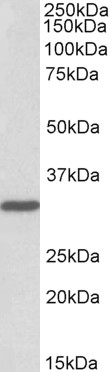
![Whole cell extract (30 μg) was separated by 12% SDS-PAGE, and the membrane was blotted with PCNA antibody [HL1966] (GTX637859) diluted at 1:1000. The HRP-conjugated anti-rabbit IgG antibody (GTX213110-01) was used to detect the primary antibody.](https://www.genetex.com/upload/website/prouct_img/normal/GTX637859/GTX637859_T-44858_20221209_WB_D_22121123_640.webp)
Product group Antibodies
PCNA antibody [HL1966]GTX637859
ApplicationsImmunoFluorescence, Western Blot, ImmunoCytoChemistry, ImmunoHistoChemistry, ImmunoHistoChemistry Paraffin
ReactivityCanine, Human
TargetPCNA
- SizePrice