

PCOLCE Antibody
LS-C750267
ApplicationsWestern Blot
Product group Antibodies
ReactivityHuman
TargetPCOLCE
Overview
- SupplierLifeSpan BioSciences
- Product NamePCOLCE Antibody
- Delivery Days Customer23
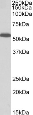
- Application Supplier NoteThe predicted MW is 47kDa, while the observed MW by Western blot was 48kDa.
- ApplicationsWestern Blot
- Applications SupplierWB (1:200 - 1:2000) The predicted MW is 47kDa, while the observed MW by Western blot was 48kDa.
- CertificationResearch Use Only
- ClonalityPolyclonal
- ConjugateUnconjugated
- Gene ID5118
- Target namePCOLCE
- Target descriptionprocollagen C-endopeptidase enhancer
- Target synonymsPCPE, PCPE-1, PCPE1, procollagen C-endopeptidase enhancer 1, procollagen C-proteinase enhancer 1, procollagen COOH-terminal proteinase enhancer 1, procollagen, type 1, COOH-terminal proteinase enhancer, type 1 procollagen C-proteinase enhancer protein, type I procollagen COOH-terminal proteinase enhancer
- HostRabbit
- IsotypeIgG
- ReactivityHuman
- Storage Instruction-20°C
- UNSPSC41116161
Related products
Product group Antibodies
Anti-PCOLCE AntibodyA85078
ApplicationsWestern Blot, ELISA
ReactivityHuman
- SizePrice

Product group Antibodies
Anti-PCOLCE Antibody Picoband(r)A06428-2-CARRIER-FREE
ApplicationsFlow Cytometry, Western Blot, ELISA, ImmunoHistoChemistry
ReactivityHuman
TargetPCOLCE
- SizePrice
Product group Antibodies

Goat anti-PCOLCEEB11815
ApplicationsWestern Blot, ELISA
ReactivityCanine, Human
TargetPCOLCE
- SizePrice
Product group Antibodies
PCOLCE AntibodyCSB-PA614881LA01HU
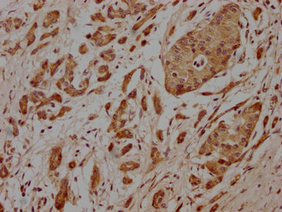
ApplicationsELISA, ImmunoHistoChemistry
ReactivityHuman
TargetPCOLCE
- SizePrice
Product group Antibodies
ApplicationsImmunoPrecipitation, Western Blot, ImmunoCytoChemistry, ImmunoHistoChemistry
ReactivityMouse
TargetPCOLCE
- SizePrice

Product group Antibodies
Anti-PCOLCE AntibodyHPA042927
ApplicationsWestern Blot, ImmunoCytoChemistry
ReactivityHuman
TargetPCOLCE
- SizePrice
![Whole cell extract (30 μg) was separated by 10% SDS-PAGE, and the membrane was blotted with PCOLCE antibody [N1C3] (GTX104154) diluted at 1:1000. The HRP-conjugated anti-rabbit IgG antibody (GTX213110-01) was used to detect the primary antibody.](https://www.genetex.com/upload/website/prouct_img/normal/GTX104154/GTX104154_43789_20191213_WB_w_23060120_931.webp)
Product group Antibodies
PCOLCE antibody [N1C3]GTX104154
ApplicationsWestern Blot, ImmunoHistoChemistry, ImmunoHistoChemistry Paraffin
ReactivityHuman
TargetPCOLCE
- SizePrice