

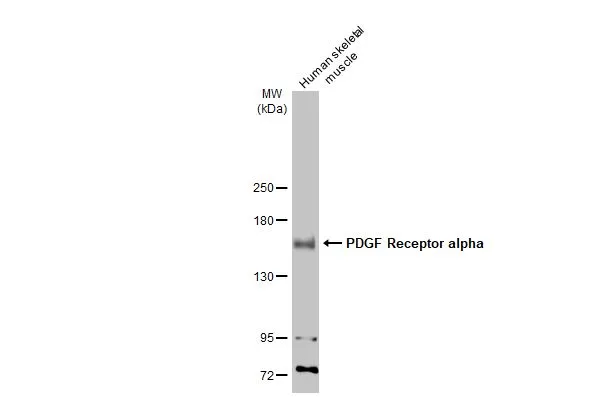
Human tissue extract (30 μg) was separated by 5% SDS-PAGE, and the membrane was blotted with PDGF Receptor alpha antibody [N2C2], Internal (GTX107903) diluted at 1:1000. The HRP-conjugated anti-rabbit IgG antibody (GTX213110-01) was used to detect the primary antibody, and the signal was developed with Trident ECL plus-Enhanced.
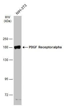
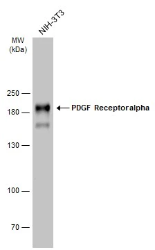
![Various whole cell extracts (30 μg) were separated by 7.5% SDS-PAGE, and the membrane was blotted with PDGF Receptor alpha antibody [N2C2], Internal (GTX107903) diluted at 1:500.](https://www.genetex.com/upload/website/prouct_img/normal/GTX107903/GTX107903_39988_20160121_WB_R_w_23060120_934.webp "Various whole cell extracts (30 μg) were separated by 7.5% SDS-PAGE, and the membrane was blotted with PDGF Receptor alpha antibody [N2C2], Internal (GTX107903) diluted at 1:500.")
 A: K562 5% SDS PAGE GTX107903 diluted at 1:1000")
![PDGF Receptor alpha antibody [N2C2], Internal detects PDGF Receptor alpha protein at cytoplasm by immunofluorescent analysis. Sample: NIH/3T3 cells were fixed in 4% paraformaldehyde at RT for 15 min. Green: PDGF Receptor alpha stained by PDGF Receptor alpha antibody [N2C2], Internal (GTX107903) diluted at 1:500. Red: phalloidin, a cytoskeleton marker diluted at 1:200. Blue: Hoechst 33342 staining.](https://www.genetex.com/upload/website/prouct_img/normal/GTX107903/GTX107903_39988_20191025_ICC_IF_w_23060120_673.webp "PDGF Receptor alpha antibody [N2C2], Internal detects PDGF Receptor alpha protein at cytoplasm by immunofluorescent analysis. Sample: NIH/3T3 cells were fixed in 4% paraformaldehyde at RT for 15 min. Green: PDGF Receptor alpha stained by PDGF Receptor alpha antibody [N2C2], Internal (GTX107903) diluted at 1:500. Red: phalloidin, a cytoskeleton marker diluted at 1:200. Blue: Hoechst 33342 staining.")
![Various whole cell extracts (30 μg) were separated by 5% SDS-PAGE, and the membrane was blotted with PDGF Receptor alpha antibody [N2C2], Internal (GTX107903) diluted at 1:2000. The HRP-conjugated anti-rabbit IgG antibody (GTX213110-01) was used to detect the primary antibody, and the signal was developed with Trident ECL plus-Enhanced.](https://www.genetex.com/upload/website/prouct_img/normal/GTX107903/GTX107903_39988_20230616_WB_M_Differentiated_23062019_816.webp "Various whole cell extracts (30 μg) were separated by 5% SDS-PAGE, and the membrane was blotted with PDGF Receptor alpha antibody [N2C2], Internal (GTX107903) diluted at 1:2000. The HRP-conjugated anti-rabbit IgG antibody (GTX213110-01) was used to detect the primary antibody, and the signal was developed with Trident ECL plus-Enhanced.")
![Various whole cell extracts (30 μg) were separated by 5% SDS-PAGE, and the membrane was blotted with PDGF Receptor alpha antibody [N2C2], Internal (GTX107903) diluted at 1:1000. The HRP-conjugated anti-rabbit IgG antibody (GTX213110-01) was used to detect the primary antibody. Corresponding RNA expression data for the same cell lines are based on Human Protein Atlas program.](https://www.genetex.com/upload/website/prouct_img/normal/GTX107903/GTX107903_45511_20240927_WB_TPM_watermark_24110700_574.webp "Various whole cell extracts (30 μg) were separated by 5% SDS-PAGE, and the membrane was blotted with PDGF Receptor alpha antibody [N2C2], Internal (GTX107903) diluted at 1:1000. The HRP-conjugated anti-rabbit IgG antibody (GTX213110-01) was used to detect the primary antibody. Corresponding RNA expression data for the same cell lines are based on Human Protein Atlas program.")
Human tissue extract (30 μg) was separated by 5% SDS-PAGE, and the membrane was blotted with PDGF Receptor alpha antibody [N2C2], Internal (GTX107903) diluted at 1:1000. The HRP-conjugated anti-rabbit IgG antibody (GTX213110-01) was used to detect the primary antibody, and the signal was developed with Trident ECL plus-Enhanced.
PDGF Receptor alpha antibody [N2C2], Internal
GTX107903
ApplicationsImmunoFluorescence, Western Blot, ImmunoCytoChemistry
Product group Antibodies
ReactivityHuman, Mouse, Rat
TargetPDGFRA
Overview
- SupplierGeneTex
- Product NamePDGF Receptor alpha antibody [N2C2], Internal
- Delivery Days Customer9
- Application Supplier NoteWB: 1:500-1:3000. *Optimal dilutions/concentrations should be determined by the researcher.Not tested in other applications.
- ApplicationsImmunoFluorescence, Western Blot, ImmunoCytoChemistry
- CertificationResearch Use Only
- ClonalityPolyclonal
- Concentration1.13 mg/ml
- ConjugateUnconjugated
- Gene ID5156
- Target namePDGFRA
- Target descriptionplatelet derived growth factor receptor alpha
- Target synonymsCD140A, PDGFR-2, PDGFR2, platelet-derived growth factor receptor alpha, CD140 antigen-like family member A, CD140a antigen, PDGF-R-alpha, alpha-type platelet-derived growth factor receptor, platelet-derived growth factor receptor 2, platelet-derived growth factor receptor, alpha polypeptide
- HostRabbit
- IsotypeIgG
- Protein IDP16234
- Protein NamePlatelet-derived growth factor receptor alpha
- Scientific DescriptionThis gene encodes a cell surface tyrosine kinase receptor for members of the platelet-derived growth factor family. These growth factors are mitogens for cells of mesenchymal origin. The identity of the growth factor bound to a receptor monomer determines whether the functional receptor is a homodimer or a heterodimer, composed of both platelet-derived growth factor receptor alpha and beta polypeptides. Studies in knockout mice, where homozygosity is lethal, indicate that the alpha form of the platelet-derived growth factor receptor is particularly important for kidney development since mice heterozygous for the receptor exhibit defective kidney phenotypes. [provided by RefSeq]
- ReactivityHuman, Mouse, Rat
- Storage Instruction-20°C or -80°C,2°C to 8°C
- UNSPSC41116161
Datasheet
Related products
Product group Antibodies
ApplicationsImmunoFluorescence, Western Blot, ELISA, ImmunoHistoChemistry
ReactivityHuman, Mouse, Rat
- SizePrice
Product group Antibodies
Anti-PDGFRA Antibody144-02103
ApplicationsWestern Blot
ReactivityHuman, Mouse, Rat
TargetPDGFRA
- SizePrice
Product group Antibodies

ApplicationsImmunoFluorescence, ImmunoHistoChemistry, ImmunoHistoChemistry Paraffin
ReactivityHuman, Mouse, Rat
TargetPDGFRA
- SizePrice

Product group Antibodies
Anti-PDGFRA Antibody Picoband(r)A00366-CARRIER-FREE
ApplicationsImmunoFluorescence, Western Blot, ELISA, ImmunoCytoChemistry
ReactivityHuman, Mouse, Rat
TargetPDGFRA
- SizePrice
Product group Antibodies
References
PDGFRA Polyclonal AntibodyBS-0231R
ApplicationsImmunoFluorescence, ELISA, ImmunoCytoChemistry, ImmunoHistoChemistry, ImmunoHistoChemistry Frozen, ImmunoHistoChemistry Paraffin
ReactivityBovine, Canine, Chicken, Equine, Human, Mouse, Porcine, Rat
TargetPDGFRA
- SizePrice
Product group Antibodies
PDGFRA AntibodyCSB-PA003724
ApplicationsImmunoFluorescence, Western Blot, ELISA, ImmunoHistoChemistry
ReactivityHuman, Mouse, Rat
TargetPDGFRA
- SizePrice
Product group Antibodies
Pdgfra Polyclonal AntibodyCAC08107
ApplicationsImmunoFluorescence, Western Blot, ELISA
ReactivityMouse
TargetPDGFRA
- SizePrice
Product group Antibodies
PDGF Receptor alpha antibodyGTX133588
ApplicationsWestern Blot
ReactivityHuman, Mouse
TargetPDGFRA
- SizePrice
Product group Antibodies
PDGF Receptor alpha antibodyGTX133619
ApplicationsImmunoFluorescence, Western Blot, ImmunoCytoChemistry, ImmunoHistoChemistry, ImmunoHistoChemistry Frozen
ReactivityHuman, Mouse
TargetPDGFRA
- SizePrice