

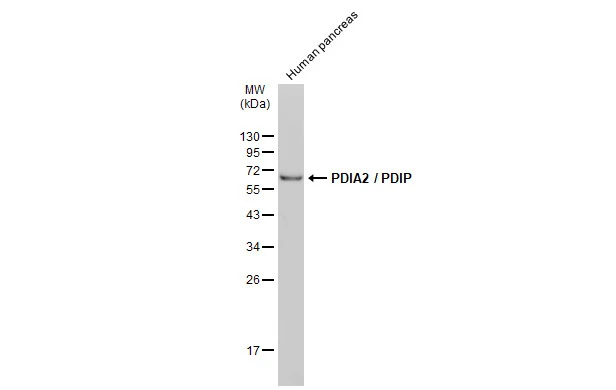
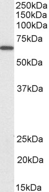
Human tissue extract (30 μg) was separated by 12% SDS-PAGE, and the membrane was blotted with PDIA2 / PDIP antibody (GTX132895) diluted at 1:500. The HRP-conjugated anti-rabbit IgG antibody (GTX213110-01) was used to detect the primary antibody.
Human tissue extract (30 μg) was separated by 12% SDS-PAGE, and the membrane was blotted with PDIA2 / PDIP antibody (GTX132895) diluted at 1:500. The HRP-conjugated anti-rabbit IgG antibody (GTX213110-01) was used to detect the primary antibody.
PDIA2 / PDIP antibody
GTX132895
ApplicationsWestern Blot
Product group Antibodies
ReactivityHuman
TargetPDIA2
Overview
- SupplierGeneTex
- Product NamePDIA2 / PDIP antibody
- Delivery Days Customer9
- Application Supplier NoteWB: 1:500-1:3000. *Optimal dilutions/concentrations should be determined by the researcher.Not tested in other applications.
- ApplicationsWestern Blot
- CertificationResearch Use Only
- ClonalityPolyclonal
- Concentration1.51 mg/ml
- ConjugateUnconjugated
- Gene ID64714
- Target namePDIA2
- Target descriptionprotein disulfide isomerase family A member 2
- Target synonymsPDA2, PDI, PDIP, PDIR, protein disulfide-isomerase A2, Rho GDP dissociation inhibitor gamma, pancreas-specific protein disulfide isomerase, pancreatic protein disulfide isomerase, protein disulfide isomerase, pancreatic, protein disulfide isomerase-associated 2
- HostRabbit
- IsotypeIgG
- Protein IDQ13087
- Protein NameProtein disulfide-isomerase A2
- Scientific DescriptionThis gene encodes a member of the disulfide isomerase (PDI) family of endoplasmic reticulum (ER) proteins that catalyze protein folding and thiol-disulfide interchange reactions. The encoded protein has an N-terminal ER-signal sequence, two catalytically active thioredoxin (TRX) domains, two TRX-like domains and a C-terminal ER-retention sequence. The protein plays a role in the folding of nascent proteins in the endoplasmic reticulum by forming disulfide bonds through its thiol isomerase, oxidase, and reductase activity. The encoded protein also possesses estradiol-binding activity and can modulate intracellular estradiol levels. [provided by RefSeq, Sep 2017]
- ReactivityHuman
- Storage Instruction-20°C or -80°C,2°C to 8°C
- UNSPSC41116161
Datasheet
Related products
Product group Antibodies
ApplicationsWestern Blot, ELISA
ReactivityHuman, Mouse, Rat
- SizePrice

Product group Antibodies
Anti-PDIP/PDIA2 Antibody Picoband(r)A03275-1-CARRIER-FREE
ApplicationsFlow Cytometry, Western Blot, ELISA
ReactivityHuman
TargetPDIA2
- SizePrice
Product group Antibodies
Anti-PDIA2 Antibody144-60103
ApplicationsWestern Blot
ReactivityHuman, Mouse, Rat
TargetPDIA2
- SizePrice
Product group Antibodies
PDIA2 Recombinant Antibody, Biotin ConjugatedBSM-61394R-BIOTIN
ApplicationsWestern Blot
ReactivityHuman, Mouse, Rat
TargetPDIA2
- SizePrice
Product group Antibodies

Goat anti-PDIA2 / PDIPEB07722
ApplicationsWestern Blot, ELISA, ImmunoHistoChemistry
ReactivityHuman, Mouse, Rat
TargetPDIA2
- SizePrice
Product group Antibodies
ApplicationsImmunoPrecipitation, Western Blot, ImmunoCytoChemistry, ImmunoHistoChemistry
ReactivityPorcine
TargetPDIA2
- SizePrice
Product group Antibodies
PDIA2 AntibodyCSB-PA269141
ApplicationsELISA, ImmunoHistoChemistry
ReactivityHuman
TargetPDIA2
- SizePrice
Product group Antibodies

PDIA2 AntibodyLS-C402749
ApplicationsELISA, ImmunoHistoChemistry
ReactivityHuman
TargetPDIA2
- SizePrice
Product group Antibodies
PDIA2 / PDIP antibody, InternalGTX89110
ApplicationsWestern Blot, ImmunoHistoChemistry, ImmunoHistoChemistry Paraffin
ReactivityHuman
TargetPDIA2
- SizePrice

Product group Antibodies
Anti-PDIA2 AntibodyHPA051692
ApplicationsImmunoHistoChemistry
ReactivityHuman
TargetPDIA2
- SizePrice