

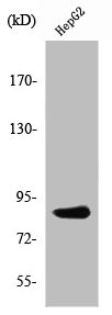
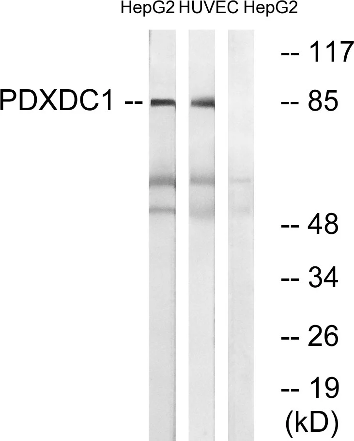
Western Blot analysis of HuvEc cells using PDXDC1 Polyclonal Antibody
Western Blot analysis of HuvEc cells using PDXDC1 Polyclonal Antibody
PDXDC1 Antibody
CSB-PA003734
ApplicationsWestern Blot, ELISA, ImmunoHistoChemistry
Product group Antibodies
ReactivityHuman, Mouse, Rat
TargetPDXDC1
Overview
- SupplierCusabio
- Product NamePDXDC1 Antibody
- Delivery Days Customer20
- ApplicationsWestern Blot, ELISA, ImmunoHistoChemistry
- CertificationResearch Use Only
- ClonalityPolyclonal
- ConjugateUnconjugated
- Gene ID23042
- Target namePDXDC1
- Target descriptionpyridoxal dependent decarboxylase domain containing 1
- Target synonymsLP8165, pyridoxal-dependent decarboxylase domain-containing protein 1
- HostRabbit
- IsotypeIgG
- Protein IDQ6P996
- Protein NamePyridoxal-dependent decarboxylase domain-containing protein 1
- ReactivityHuman, Mouse, Rat
- Storage Instruction-20°C or -80°C
- UNSPSC41116161
Related products

Product group Antibodies
Anti-PDXDC1 AntibodyA96796
ApplicationsWestern Blot, ELISA
ReactivityHuman, Mouse, Rat
- SizePrice

Product group Antibodies
Anti-PDXDC1 Antibody Picoband(r)A11375-2-CARRIER-FREE
ApplicationsFlow Cytometry, Western Blot, ELISA, ImmunoHistoChemistry
ReactivityHuman, Mouse, Rat
TargetPDXDC1
- SizePrice
Product group Antibodies
Anti-PDXDC1 Antibody144-60825
ApplicationsWestern Blot
ReactivityHuman, Mouse, Rat
TargetPDXDC1
- SizePrice
Product group Antibodies

PDXDC1 AntibodyLS-C760992
ApplicationsWestern Blot, ImmunoHistoChemistry
ReactivityHuman
TargetPDXDC1
- SizePrice
Product group Antibodies
PDXDC1 antibodyGTX87408
ApplicationsWestern Blot
ReactivityHuman
TargetPDXDC1
- SizePrice

Product group Antibodies
Anti-PDXDC1 AntibodyHPA049121
ApplicationsImmunoCytoChemistry
ReactivityHuman
TargetPDXDC1
- SizePrice
Product group Antibodies
Anti-PDXDC1Y058742
ApplicationsWestern Blot, ELISA
ReactivityHuman, Mouse, Rat
- SizePrice