

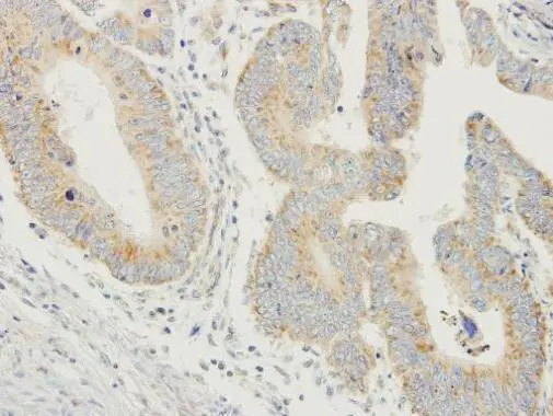
Immunohistochemical analysis of paraffin-embedded human colon carcinoma, using PDYN(GTX113515) antibody at 1:250 dilution.
Antigen Retrieval: Trilogy? (EDTA based, pH 8.0) buffer, 15min
![Non-transfected (–) and transfected (+) 293T whole cell extracts (30 μg) were separated by 12% SDS-PAGE, and the membrane was blotted with ProDynorphin antibody [N1C2] (GTX113515) diluted at 1:5000. The HRP-conjugated anti-rabbit IgG antibody (GTX213110-01) was used to detect the primary antibody.](https://www.genetex.com/upload/website/prouct_img/normal/GTX113515/GTX113515_45105_20240809_WB_B_24082301_579.webp "Non-transfected (–) and transfected (+) 293T whole cell extracts (30 μg) were separated by 12% SDS-PAGE, and the membrane was blotted with ProDynorphin antibody [N1C2] (GTX113515) diluted at 1:5000. The HRP-conjugated anti-rabbit IgG antibody (GTX213110-01) was used to detect the primary antibody.")
![Human tissue extract (5 μg) was separated by 12% SDS-PAGE, and the membrane was blotted with ProDynorphin antibody [N1C2] (GTX113515) diluted at 1:500. The HRP-conjugated anti-rabbit IgG antibody (GTX213110-01) was used to detect the primary antibody, and the signal was developed with Trident ECL plus-Enhanced.](https://www.genetex.com/upload/website/prouct_img/normal/GTX113515/GTX113515_45105_20240809_WB_brain_24082301_244.webp "Human tissue extract (5 μg) was separated by 12% SDS-PAGE, and the membrane was blotted with ProDynorphin antibody [N1C2] (GTX113515) diluted at 1:500. The HRP-conjugated anti-rabbit IgG antibody (GTX213110-01) was used to detect the primary antibody, and the signal was developed with Trident ECL plus-Enhanced.")
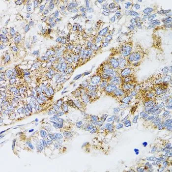
Immunohistochemical analysis of paraffin-embedded human colon carcinoma, using PDYN(GTX113515) antibody at 1:250 dilution.
Antigen Retrieval: Trilogy? (EDTA based, pH 8.0) buffer, 15min
ProDynorphin antibody [N1C2]
GTX113515
ApplicationsWestern Blot, ImmunoHistoChemistry, ImmunoHistoChemistry Paraffin
Product group Antibodies
ReactivityHuman
TargetPDYN
Overview
- SupplierGeneTex
- Product NameProDynorphin antibody [N1C2]
- Delivery Days Customer9
- Application Supplier NoteWB: 1:500-1:3000. IHC-P: 1:100-1:1000. *Optimal dilutions/concentrations should be determined by the researcher.Not tested in other applications.
- ApplicationsWestern Blot, ImmunoHistoChemistry, ImmunoHistoChemistry Paraffin
- CertificationResearch Use Only
- ClonalityPolyclonal
- Concentration1.23 mg/ml
- ConjugateUnconjugated
- Gene ID5173
- Target namePDYN
- Target descriptionprodynorphin
- Target synonymsADCA, PENKB, SCA23, proenkephalin-B, beta-neoendorphin-dynorphin, leu-enkephalin, leumorphin, neoendorphin-dynorphin-enkephalin prepropeptide, preprodynorphin, preproenkephalin B, rimorphin
- HostRabbit
- IsotypeIgG
- Protein IDP01213
- Protein NameProenkephalin-B
- Scientific DescriptionThe protein encoded by this gene is a preproprotein that is proteolytically processed to form the secreted opioid peptides beta-neoendorphin, dynorphin, leu-enkephalin, rimorphin, and leumorphin. These peptides are ligands for the kappa-type of opioid receptor. Dynorphin is involved in modulating responses to several psychoactive substances, including cocaine. [provided by RefSeq]
- ReactivityHuman
- Storage Instruction-20°C or -80°C,2°C to 8°C
- UNSPSC41116161
Datasheet
Related products
Product group Antibodies
ApplicationsImmunoPrecipitation, Western Blot, ImmunoCytoChemistry, ImmunoHistoChemistry
TargetPDYN
- SizePrice
Product group Antibodies
Anti-Dynorphin A Antibody130-10426
ApplicationsWestern Blot, ELISA
TargetPDYN
- SizePrice
Product group Antibodies
Anti-Dynorphin A AntibodyA283838
ApplicationsImmunoHistoChemistry, ImmunoHistoChemistry Frozen, ImmunoHistoChemistry Paraffin, RadioImmunoAssay
ReactivityHuman, Porcine, Primate, Rat
- SizePrice
Product group Antibodies
ApplicationsWestern Blot, ImmunoHistoChemistry
ReactivityHuman, Mouse, Rat
TargetPDYN
- SizePrice
Product group Antibodies

ApplicationsWestern Blot, ELISA
ReactivityHuman
TargetPDYN
- SizePrice

Product group Antibodies
Anti-PDYN AntibodyHPA053342
ApplicationsImmunoHistoChemistry
ReactivityHuman
TargetPDYN
- SizePrice
![Non-transfected (–) and transfected (+) 293T whole cell extracts (30 μg) were separated by 12% SDS-PAGE, and the membrane was blotted with ProDynorphin antibody [HL2573] (GTX638951) diluted at 1:50000. The HRP-conjugated anti-rabbit IgG antibody (GTX213110-01) was used to detect the primary antibody.](https://www.genetex.com/upload/website/prouct_img/normal/GTX638951/GTX638951_T-45152_20240816_WB_B_24082301_167.webp)
Product group Antibodies
ProDynorphin antibody [HL2573]GTX638951
ApplicationsWestern Blot
ReactivityHuman
TargetPDYN
- SizePrice
![PDYN antibody [HL2574] detects secreted PDYN protein by immunohistochemical analysis. Sample: Paraffin-embedded human glioblastoma. PDYN stained by PDYN antibody [HL2574] (GTX638952) diluted at 1:100. Antigen Retrieval: Citrate buffer, pH 6.0, 15 min](https://www.genetex.com/upload/website/prouct_img/normal/GTX638952/GTX638952_T-45152_20230915_IHC-P_24021917_544.webp)
Product group Antibodies
ProDynorphin antibody [HL2574]GTX638952
ApplicationsWestern Blot, ImmunoHistoChemistry, ImmunoHistoChemistry Paraffin
ReactivityHuman
TargetPDYN
- SizePrice
![Various tissue extracts (50 μg) were separated by 12% SDS-PAGE, and the membrane was blotted with ProDynorphin antibody [HL3295] (GTX640984) diluted at 1:1000. The HRP-conjugated anti-rabbit IgG antibody (GTX213110-01) was used to detect the primary antibody.](https://www.genetex.com/upload/website/prouct_img/normal/GTX640984/GTX640984_T-45537_20241004_WB_M_R_24100900_462.webp)
Product group Antibodies
ProDynorphin antibody [HL3295]GTX640984
ApplicationsWestern Blot
ReactivityHuman, Mouse, Rat
TargetPDYN
- SizePrice
Product group Antibodies
ProDynorphin antibodyGTX65945
ApplicationsWestern Blot, ImmunoHistoChemistry, ImmunoHistoChemistry Paraffin
ReactivityHuman, Mouse, Rat
TargetPDYN
- SizePrice