

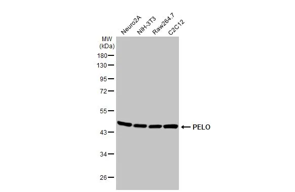
Various whole cell extracts (30 μg) were separated by 10% SDS-PAGE, and the membrane was blotted with PELO antibody [HL2345] (GTX638548) diluted at 1:1000. The HRP-conjugated anti-rabbit IgG antibody (GTX213110-01) was used to detect the primary antibody.
![Various whole cell extracts (30 μg) were separated by 10% SDS-PAGE, and the membrane was blotted with PELO antibody [HL2345] (GTX638548) diluted at 1:1000. The HRP-conjugated anti-rabbit IgG antibody (GTX213110-01) was used to detect the primary antibody.](https://www.genetex.com/upload/website/prouct_img/normal/GTX638548/GTX638548_T-45026_20230428_WB_R_23050223_522.webp "Various whole cell extracts (30 μg) were separated by 10% SDS-PAGE, and the membrane was blotted with PELO antibody [HL2345] (GTX638548) diluted at 1:1000. The HRP-conjugated anti-rabbit IgG antibody (GTX213110-01) was used to detect the primary antibody.")
![PELO antibody [HL2345] detects PELO protein by immunohistochemical analysis. Sample: Paraffin-embedded rat tissues. PELO stained by PELO antibody [HL2345] (GTX638548) diluted at 1:100. Antigen Retrieval: Citrate buffer, pH 6.0, 15 min](https://www.genetex.com/upload/website/prouct_img/normal/GTX638548/GTX638548_T-45026_20230512_IHC-P_multiple_R_23060622_924.webp "PELO antibody [HL2345] detects PELO protein by immunohistochemical analysis. Sample: Paraffin-embedded rat tissues. PELO stained by PELO antibody [HL2345] (GTX638548) diluted at 1:100. Antigen Retrieval: Citrate buffer, pH 6.0, 15 min")
![PELO antibody [HL2345] detects PELO protein at cytoplasm and nucleus by immunohistochemical analysis. Sample: Paraffin-embedded human hepatocellular carcinoma. PELO stained by PELO antibody [HL2345] (GTX638548) diluted at 1:100. Antigen Retrieval: Citrate buffer, pH 6.0, 15 min](https://www.genetex.com/upload/website/prouct_img/normal/GTX638548/GTX638548_T-45026_20230512_IHC-P_23060622_698.webp "PELO antibody [HL2345] detects PELO protein at cytoplasm and nucleus by immunohistochemical analysis. Sample: Paraffin-embedded human hepatocellular carcinoma. PELO stained by PELO antibody [HL2345] (GTX638548) diluted at 1:100. Antigen Retrieval: Citrate buffer, pH 6.0, 15 min")
![PELO antibody [HL2345] detects PELO protein by immunohistochemical analysis. Sample: Paraffin-embedded mouse tissues. PELO stained by PELO antibody [HL2345] (GTX638548) diluted at 1:100. Antigen Retrieval: Citrate buffer, pH 6.0, 15 min](https://www.genetex.com/upload/website/prouct_img/normal/GTX638548/GTX638548_T-45026_20230512_IHC-P_multiple_M_23060622_504.webp "PELO antibody [HL2345] detects PELO protein by immunohistochemical analysis. Sample: Paraffin-embedded mouse tissues. PELO stained by PELO antibody [HL2345] (GTX638548) diluted at 1:100. Antigen Retrieval: Citrate buffer, pH 6.0, 15 min")
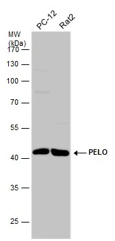
![Various whole cell extracts (30 μg) were separated by 10% SDS-PAGE, and the membrane was blotted with PELO antibody [HL2345] (GTX638548) diluted at 1:1000. The HRP-conjugated anti-rabbit IgG antibody (GTX213110-01) was used to detect the primary antibody.](https://www.genetex.com/upload/website/prouct_img/normal/GTX638548/GTX638548_45125_20230811_WB_23081619_265.webp "Various whole cell extracts (30 μg) were separated by 10% SDS-PAGE, and the membrane was blotted with PELO antibody [HL2345] (GTX638548) diluted at 1:1000. The HRP-conjugated anti-rabbit IgG antibody (GTX213110-01) was used to detect the primary antibody.")
![Whole zebrafish extract (30 μg) was separated by 10% SDS-PAGE, and the membrane was blotted with PELO antibody [HL2345] (GTX638548) diluted at 1:1000. The HRP-conjugated anti-rabbit IgG antibody (GTX213110-01) was used to detect the primary antibody.](https://www.genetex.com/upload/website/prouct_img/normal/GTX638548/GTX638548_45125_20230811_WB_Z_23081619_776.webp "Whole zebrafish extract (30 μg) was separated by 10% SDS-PAGE, and the membrane was blotted with PELO antibody [HL2345] (GTX638548) diluted at 1:1000. The HRP-conjugated anti-rabbit IgG antibody (GTX213110-01) was used to detect the primary antibody.")
![PELO antibody [HL2345] detects PELO protein on whole mount zebrafish by immunohistochemical analysis. Sample: Paraformaldehyde-fixed 2 days-post-fertilization zebrafish embryo. Green: PELO stained by PELO antibody [HL2345] (GTX638548) diluted at 1:100. Antigen Retrieval: Tris-HCl buffer, pH 9.0, 20 min at 70oC](https://www.genetex.com/upload/website/prouct_img/normal/GTX638548/GTX638548_45125_20230928_IHC-Wm_Z_23100319_567.webp "PELO antibody [HL2345] detects PELO protein on whole mount zebrafish by immunohistochemical analysis. Sample: Paraformaldehyde-fixed 2 days-post-fertilization zebrafish embryo. Green: PELO stained by PELO antibody [HL2345] (GTX638548) diluted at 1:100. Antigen Retrieval: Tris-HCl buffer, pH 9.0, 20 min at 70oC")
![PELO antibody [HL2345] detects PELO protein on whole mount zebrafish by immunohistochemical analysis. Sample: Paraformaldehyde-fixed 2 days-post-fertilization zebrafish embryo. Green: PELO stained by PELO antibody [HL2345] (GTX638548) diluted at 1:100. Antigen Retrieval: Tris-HCl buffer, pH 9.0, 20 min at 70oC](https://www.genetex.com/upload/website/prouct_img/normal/GTX638548/GTX638548_45125_20230928_IHC-Wm_Z_1_23100319_966.webp "PELO antibody [HL2345] detects PELO protein on whole mount zebrafish by immunohistochemical analysis. Sample: Paraformaldehyde-fixed 2 days-post-fertilization zebrafish embryo. Green: PELO stained by PELO antibody [HL2345] (GTX638548) diluted at 1:100. Antigen Retrieval: Tris-HCl buffer, pH 9.0, 20 min at 70oC")
![Whole Japanese medaka extract (30 μg) was separated by 10% SDS-PAGE, and the membrane was blotted with PELO antibody [HL2345] (GTX638548) diluted at 1:1000. The HRP-conjugated anti-rabbit IgG antibody (GTX213110-01) was used to detect the primary antibody.](https://www.genetex.com/upload/website/prouct_img/normal/GTX638548/GTX638548_45125_20250815_WB_medaka_25082121_214.webp "Whole Japanese medaka extract (30 μg) was separated by 10% SDS-PAGE, and the membrane was blotted with PELO antibody [HL2345] (GTX638548) diluted at 1:1000. The HRP-conjugated anti-rabbit IgG antibody (GTX213110-01) was used to detect the primary antibody.")
Various whole cell extracts (30 μg) were separated by 10% SDS-PAGE, and the membrane was blotted with PELO antibody [HL2345] (GTX638548) diluted at 1:1000. The HRP-conjugated anti-rabbit IgG antibody (GTX213110-01) was used to detect the primary antibody.
PELO antibody [HL2345]
GTX638548
ApplicationsWestern Blot, ImmunoHistoChemistry, ImmunoHistoChemistry Paraffin
Product group Antibodies
ReactivityHuman, Mouse, Rat, Zebra Fish
TargetPELO
Overview
- SupplierGeneTex
- Product NamePELO antibody [HL2345]
- Delivery Days Customer9
- Application Supplier NoteWB: 1:500-1:3000. *Optimal dilutions/concentrations should be determined by the researcher.Not tested in other applications.
- ApplicationsWestern Blot, ImmunoHistoChemistry, ImmunoHistoChemistry Paraffin
- CertificationResearch Use Only
- ClonalityMonoclonal
- Clone IDHL2345
- Concentration1 mg/ml
- ConjugateUnconjugated
- Gene ID53918
- Target namePELO
- Target descriptionpelota mRNA surveillance and ribosome rescue factor
- Target synonymsCGI-17, PRO1770, protein pelota homolog, hPelota, pelota homolog, protein Dom34 homolog
- HostRabbit
- IsotypeIgG
- Protein IDQ9BRX2
- Protein NameProtein pelota homolog
- Scientific DescriptionThis gene encodes a protein which contains a conserved nuclear localization signal. The encoded protein may have a role in spermatogenesis, cell cycle control, and in meiotic cell division. [provided by RefSeq, Jul 2008]
- ReactivityHuman, Mouse, Rat, Zebra Fish
- Storage Instruction-20°C or -80°C,2°C to 8°C
- UNSPSC41116161
Datasheet
Related products
Product group Antibodies
Anti-PELO AntibodyA55057
ApplicationsDot Blot, ImmunoPrecipitation, Western Blot, ELISA, ImmunoHistoChemistry
ReactivityHuman, Monkey, Mouse, Rat
- SizePrice
Product group Antibodies
PELO Polyclonal AntibodyBS-7821R
ApplicationsImmunoFluorescence, Western Blot, ELISA, ImmunoCytoChemistry, ImmunoHistoChemistry, ImmunoHistoChemistry Frozen, ImmunoHistoChemistry Paraffin
ReactivityBovine, Canine, Chicken, Equine, Human, Mouse, Rabbit, Rat
TargetPELO
- SizePrice
Product group Antibodies
PELO AntibodyCSB-PA861124LA01HU
ApplicationsELISA, ImmunoHistoChemistry
ReactivityHuman
TargetPELO
- SizePrice
Product group Antibodies

PELO AntibodyLS-C379263
ApplicationsELISA, ImmunoHistoChemistry, ImmunoHistoChemistry Paraffin
ReactivityHuman
TargetPELO
- SizePrice
Product group Antibodies
PELO antibodyGTX120446
ApplicationsImmunoFluorescence, Western Blot, ImmunoCytoChemistry, ImmunoHistoChemistry, ImmunoHistoChemistry Paraffin
ReactivityHuman, Mouse, Rat
TargetPELO
- SizePrice

Product group Antibodies
Anti-PELO AntibodyHPA031458
ApplicationsImmunoCytoChemistry, ImmunoHistoChemistry
ReactivityHuman
TargetPELO
- SizePrice
Product group Antibodies
Anti-PELOY058747
ApplicationsWestern Blot, ELISA, ImmunoHistoChemistry
ReactivityHuman, Mouse, Rat
- SizePrice