

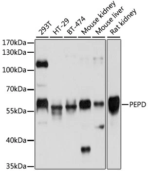
WB analysis of various sample lysates using GTX54636 PEPD antibody. Dilution : 1:1000 Loading : 25μg per lane
WB analysis of various sample lysates using GTX54636 PEPD antibody. Dilution : 1:1000 Loading : 25μg per lane
PEPD antibody
GTX54636
ApplicationsWestern Blot
Product group Antibodies
ReactivityHuman, Mouse, Rat
TargetPEPD
Overview
- SupplierGeneTex
- Product NamePEPD antibody
- Delivery Days Customer7
- Application Supplier NoteWB: 1:500 - 1:2000. *Optimal dilutions/concentrations should be determined by the researcher.Not tested in other applications.
- ApplicationsWestern Blot
- CertificationResearch Use Only
- ClonalityPolyclonal
- ConjugateUnconjugated
- Gene ID5184
- Target namePEPD
- Target descriptionpeptidase D
- Target synonymsPROLIDASE, xaa-Pro dipeptidase, X-Pro dipeptidase, aminoacyl-L-proline hydrolase, imidodipeptidase, proline dipeptidase, testicular tissue protein Li 138
- HostRabbit
- IsotypeIgG
- Protein IDP12955
- Protein NameXaa-Pro dipeptidase
- Scientific DescriptionThis gene encodes a member of the peptidase family. The protein forms a homodimer that hydrolyzes dipeptides or tripeptides with C-terminal proline or hydroxyproline residues. The enzyme serves an important role in the recycling of proline, and may be rate limiting for the production of collagen. Mutations in this gene result in prolidase deficiency, which is characterized by the excretion of large amount of di- and tri-peptides containing proline. Multiple transcript variants encoding different isoforms have been found for this gene.[provided by RefSeq, Oct 2009]
- ReactivityHuman, Mouse, Rat
- Storage Instruction-20°C or -80°C,2°C to 8°C
- UNSPSC41116161
Datasheet
Related products
Product group Antibodies
Anti-PEPD AntibodyA30778
ApplicationsWestern Blot, ImmunoHistoChemistry
ReactivityHuman, Mouse, Rat
- SizePrice

Product group Antibodies
Anti-PEPD Antibody Picoband(r)A03417-1-CARRIER-FREE
ApplicationsFlow Cytometry, ImmunoFluorescence, Western Blot, ELISA, ImmunoCytoChemistry
ReactivityHuman, Mouse, Rat
TargetPEPD
- SizePrice
Product group Antibodies
Anti-PEPD Antibody144-05416
ApplicationsWestern Blot
ReactivityHuman, Mouse, Rat
TargetPEPD
- SizePrice
Product group Antibodies

PEPD / PROLIDASE AntibodyLS-C830818
ApplicationsWestern Blot, ELISA, ImmunoHistoChemistry
ReactivityHuman, Mouse, Rat
TargetPEPD
- SizePrice
Product group Antibodies
ApplicationsFlow Cytometry, Western Blot, ImmunoCytoChemistry
ReactivityHuman
TargetPEPD
- SizePrice
Product group Antibodies
PEPD Polyclonal AntibodyCAC14748
ApplicationsWestern Blot, ELISA, ImmunoHistoChemistry
TargetPEPD
- SizePrice
Product group Antibodies
PEPD AntibodyCSB-PA017784LA01HU
ApplicationsWestern Blot, ELISA, ImmunoHistoChemistry
ReactivityHuman
TargetPEPD
- SizePrice
![WB analysis of human tissues (Lane 1-Testis ; Lane 2-Omentum ; Lane 3-Uterus ; Lane 4-Breast ; Lane 5-Brain ; Lane 6-Liver ; Lane 7-Ovary ; Lane 8-Thyroid gland ; Lane 9-colon ; Lane 10-spleen) using GTX83909 PEPD antibody [1B7]. Loading : 10 ug per lane Dilution : 1:200](https://www.genetex.com/upload/website/prouct_img/normal/GTX83909/GTX83909_3781_WB_w_23061420_321.webp)
Product group Antibodies
PEPD antibody [1B7]GTX83909
ApplicationsFlow Cytometry, ImmunoFluorescence, Western Blot, ImmunoCytoChemistry, ImmunoHistoChemistry, ImmunoHistoChemistry Paraffin
ReactivityHuman, Mouse
TargetPEPD
- SizePrice
![IHC-P analysis of human prostate tissue using GTX83910 PEPD antibody [5D5].](https://www.genetex.com/upload/website/prouct_img/normal/GTX83910/GTX83910_2012_IHC-P_w_23061420_182.webp)
Product group Antibodies
PEPD antibody [5D5]GTX83910
ApplicationsFlow Cytometry, Western Blot, ImmunoHistoChemistry, ImmunoHistoChemistry Paraffin
ReactivityCanine, Human, Mouse, Rat
TargetPEPD
- SizePrice

Product group Antibodies
Anti-PEPD AntibodyHPA072045
ApplicationsImmunoCytoChemistry
ReactivityHuman
TargetPEPD
- SizePrice