

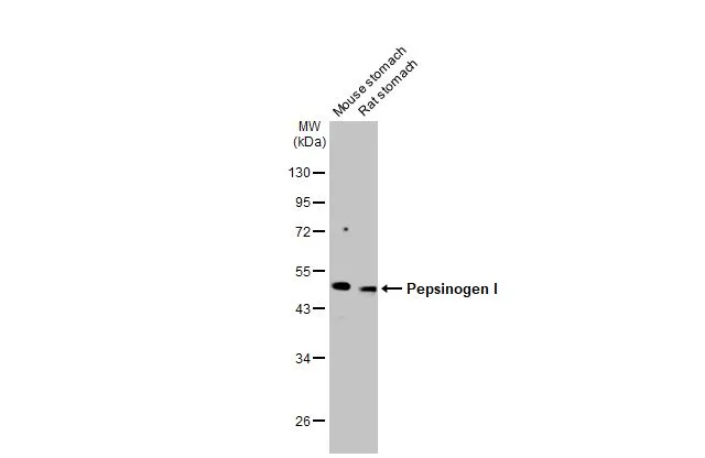
Various tissue extracts (50 μg) were separated by 10% SDS-PAGE, and the membrane was blotted with Pepsinogen I antibody [HL2137] (GTX638109) diluted at 1:1000. The HRP-conjugated anti-rabbit IgG antibody (GTX213110-01) was used to detect the primary antibody, and the signal was developed with Trident ECL plus-Enhanced.
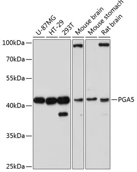
![Various tissue extracts (10 μg) were separated by 10% SDS-PAGE, and the membrane was blotted with Pepsinogen I antibody [HL2137] (GTX638109) diluted at 1:1000. The HRP-conjugated anti-rabbit IgG antibody (GTX213110-01) was used to detect the primary antibody.](https://www.genetex.com/upload/website/prouct_img/normal/GTX638109/GTX638109_T-44914_20230203_WB_tissue_23020621_454.webp "Various tissue extracts (10 μg) were separated by 10% SDS-PAGE, and the membrane was blotted with Pepsinogen I antibody [HL2137] (GTX638109) diluted at 1:1000. The HRP-conjugated anti-rabbit IgG antibody (GTX213110-01) was used to detect the primary antibody.")
![Pepsinogen I antibody [HL2137] detects Pepsinogen I protein at cytoplasm by immunofluorescent analysis. Sample: THP-1 cells were fixed in 4% paraformaldehyde at RT for 15 min. Green: Pepsinogen I stained by Pepsinogen I antibody [HL2137] (GTX638109) diluted at 1:500. Blue: Fluoroshield with DAPI (GTX30920).](https://www.genetex.com/upload/website/prouct_img/normal/GTX638109/GTX638109_T-44914_20230331_ICC_IF_23041023_313.webp "Pepsinogen I antibody [HL2137] detects Pepsinogen I protein at cytoplasm by immunofluorescent analysis. Sample: THP-1 cells were fixed in 4% paraformaldehyde at RT for 15 min. Green: Pepsinogen I stained by Pepsinogen I antibody [HL2137] (GTX638109) diluted at 1:500. Blue: Fluoroshield with DAPI (GTX30920).")
![Indirect ELISA analysis was performed by coating the plate with recombinant NS0 cells expressed, full-length human pepsinogen A protein (97.56-1.52 nM). Coated protein was probed with Pepsinogen I antibody [HL2137] (GTX638109) (1 μg/mL). Goat anti-rabbit IgG antibody (HRP) (GTX213110-01) (1:10000) was used to detect the bound primary antibody.](https://www.genetex.com/upload/website/prouct_img/normal/GTX638109/GTX638109_45005_20230602_ELISA_Indirect_23060622_939.webp "Indirect ELISA analysis was performed by coating the plate with recombinant NS0 cells expressed, full-length human pepsinogen A protein (97.56-1.52 nM). Coated protein was probed with Pepsinogen I antibody [HL2137] (GTX638109) (1 μg/mL). Goat anti-rabbit IgG antibody (HRP) (GTX213110-01) (1:10000) was used to detect the bound primary antibody.")
![Pepsinogen I antibody [HL2137] detects Pepsinogen I protein at cytoplasm by immunohistochemical analysis. Sample: Paraffin-embedded human stomach. Pepsinogen I stained by Pepsinogen I antibody [HL2137] (GTX638109) diluted at 1:500. Antigen Retrieval: Citrate buffer, pH 6.0, 15 min](https://www.genetex.com/upload/website/prouct_img/normal/GTX638109/GTX638109_45005_20230602_IHC-P_23061320_230.webp "Pepsinogen I antibody [HL2137] detects Pepsinogen I protein at cytoplasm by immunohistochemical analysis. Sample: Paraffin-embedded human stomach. Pepsinogen I stained by Pepsinogen I antibody [HL2137] (GTX638109) diluted at 1:500. Antigen Retrieval: Citrate buffer, pH 6.0, 15 min")
Various tissue extracts (50 μg) were separated by 10% SDS-PAGE, and the membrane was blotted with Pepsinogen I antibody [HL2137] (GTX638109) diluted at 1:1000. The HRP-conjugated anti-rabbit IgG antibody (GTX213110-01) was used to detect the primary antibody, and the signal was developed with Trident ECL plus-Enhanced.
Pepsinogen I antibody [HL2137]
GTX638109
ApplicationsImmunoFluorescence, Western Blot, ELISA, ImmunoCytoChemistry, ImmunoHistoChemistry, ImmunoHistoChemistry Paraffin
Product group Antibodies
ReactivityHuman, Mouse, Rat
TargetPGA5
Overview
- SupplierGeneTex
- Product NamePepsinogen I antibody [HL2137]
- Delivery Days Customer9
- Application Supplier NoteWB: 1:500-1:3000. *Optimal dilutions/concentrations should be determined by the researcher.Not tested in other applications.
- ApplicationsImmunoFluorescence, Western Blot, ELISA, ImmunoCytoChemistry, ImmunoHistoChemistry, ImmunoHistoChemistry Paraffin
- CertificationResearch Use Only
- ClonalityMonoclonal
- Clone IDHL2137
- Concentration1 mg/ml
- ConjugateUnconjugated
- Gene ID5222
- Target namePGA5
- Target descriptionpepsinogen A5
- Target synonymsPg5, pepsin A-5, Pepsin A-4, Pepsinogen-4, pepsin A, pepsinogen 5, group I (pepsinogen A), pepsinogen-5
- HostRabbit
- IsotypeIgG
- Protein IDP0DJD7
- Protein NamePepsin A-4
- ReactivityHuman, Mouse, Rat
- Storage Instruction-20°C or -80°C,2°C to 8°C
- UNSPSC41116161
Datasheet
Related products

Product group Antibodies
References
ApplicationsImmunoFluorescence, Western Blot, ELISA, ImmunoCytoChemistry, ImmunoHistoChemistry
ReactivityHuman, Mouse, Rat
TargetPGA5
- SizePrice
Product group Antibodies
Anti-PGA5 (116-135) Antibody130-10573
ApplicationsELISA
ReactivityHuman
TargetPGA5
- SizePrice
Product group Antibodies

PGA5 / Pepsin A AntibodyLS-C748020
ApplicationsWestern Blot
ReactivityHuman, Mouse, Rat
TargetPGA5
- SizePrice

Product group Antibodies
Anti-PGA5 AntibodyA89848
ApplicationsWestern Blot
ReactivityHuman, Mouse, Rat
- SizePrice
Product group Antibodies
PGA3/PGA4/PGA5 AntibodyCSB-PA030042
ApplicationsWestern Blot, ELISA, ImmunoHistoChemistry
ReactivityHuman
TargetPGA5
- SizePrice
Product group Antibodies
ApplicationsFlow Cytometry
TargetPGA5
- SizePrice
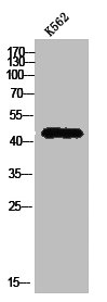
![WB analysis of HepG2 (1) and SMMC-7721 (2) cell lysate using GTX83306 PGA5 antibody [2C1].](https://www.genetex.com/upload/website/prouct_img/normal/GTX83306/GTX83306_20170912_WB_w_23061322_765.webp)
Product group Antibodies
PGA5 antibody [2C1]GTX83306
ApplicationsWestern Blot, ELISA, ImmunoHistoChemistry, ImmunoHistoChemistry Paraffin
ReactivityHuman
TargetPGA5
- SizePrice
![IHC-P analysis of human gastric antrum tissue using GTX639942 Pepsinogen I antibody [HMV318] HistoMAX?. Pepsinogen I positivity in few glands formed by chief cells in the corpus-antrum transition zone.](https://www.genetex.com/upload/website/prouct_img/normal/GTX639942/GTX639942_20240522_IHC-P_1_24052200_588.webp)
Product group Antibodies
ApplicationsImmunoHistoChemistry, ImmunoHistoChemistry Paraffin
ReactivityHuman
TargetPGA5
- SizePrice
Product group Antibodies
PGA5 antibodyGTX66144
ApplicationsWestern Blot
ReactivityHuman, Mouse, Rat
TargetPGA5
- SizePrice