

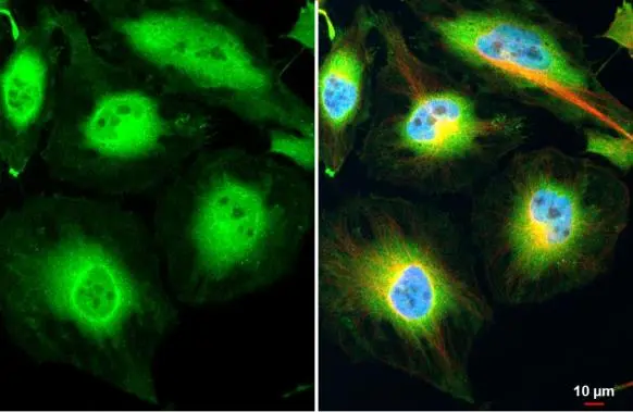
PER2 antibody detects PER2 protein at cytoplasm and nucleus by immunofluorescent analysis. Sample: HeLa cells were fixed in 4% paraformaldehyde at RT for 15 min. Green: PER2 stained by PER2 antibody (GTX129688) diluted at 1:500. Red: alpha Tubulin, a cytoskeleton marker, stained by alpha Tubulin antibody [GT114] (GTX628802) diluted at 1:1000. Blue: Fluoroshield with DAPI (GTX30920).
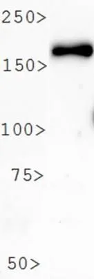
 and transfected (+) 293T whole cell extracts (30 μg) were separated by 5% SDS-PAGE, and the membrane was blotted with PER2 antibody (GTX129688) diluted at 1:20000. The HRP-conjugated anti-rabbit IgG antibody (GTX213110-01) was used to detect the primary antibody.")
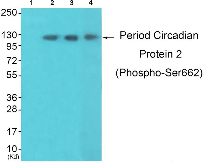
![Treated 293T whole cell extracts (30 μg) were separated by 7.5% SDS-PAGE, and the membranes were blotted with PER2 antibody (GTX129688) and BMAL1 antibody [N1N3] (GTX105060) diluted at 1:500. The HRP-conjugated anti-rabbit IgG antibody (GTX213110-01) was used to detect the primary antibody.](https://www.genetex.com/upload/website/prouct_img/normal/GTX129688/GTX129688_41605_20191025_WB_treatment_Serumshock_watermark_w_23060523_629.webp "Treated 293T whole cell extracts (30 μg) were separated by 7.5% SDS-PAGE, and the membranes were blotted with PER2 antibody (GTX129688) and BMAL1 antibody [N1N3] (GTX105060) diluted at 1:500. The HRP-conjugated anti-rabbit IgG antibody (GTX213110-01) was used to detect the primary antibody.")
 and transfected (+) 293T whole cell extracts (30 μg) were separated by 5% SDS-PAGE, and the membrane was blotted with PER2 antibody (GTX129688) diluted at 1:500. The HRP-conjugated anti-rabbit IgG antibody (GTX213110-01) was used to detect the primary antibody, and the signal was developed with Trident ECL plus-Enhanced.")
 were separated by 5% SDS-PAGE, and the membrane was blotted with PER2 antibody (GTX129688) diluted at 1:500. The HRP-conjugated anti-rabbit IgG antibody (GTX213110-01) was used to detect the primary antibody.")
PER2 antibody detects PER2 protein at cytoplasm and nucleus by immunofluorescent analysis. Sample: HeLa cells were fixed in 4% paraformaldehyde at RT for 15 min. Green: PER2 stained by PER2 antibody (GTX129688) diluted at 1:500. Red: alpha Tubulin, a cytoskeleton marker, stained by alpha Tubulin antibody [GT114] (GTX628802) diluted at 1:1000. Blue: Fluoroshield with DAPI (GTX30920).
PER2 antibody
GTX129688
ApplicationsImmunoFluorescence, Western Blot, ImmunoCytoChemistry
Product group Antibodies
ReactivityHuman
TargetPER2
Overview
- SupplierGeneTex
- Product NamePER2 antibody
- Delivery Days Customer9
- Application Supplier NoteWB: 1:500-1:20000. *Optimal dilutions/concentrations should be determined by the researcher.Not tested in other applications.
- ApplicationsImmunoFluorescence, Western Blot, ImmunoCytoChemistry
- CertificationResearch Use Only
- ClonalityPolyclonal
- Concentration1.48 mg/ml
- ConjugateUnconjugated
- Gene ID8864
- Target namePER2
- Target descriptionperiod circadian regulator 2
- Target synonymsFASPS, FASPS1, period circadian protein homolog 2, circadian clock protein PERIOD 2, hPER2, period 2, period circadian clock 2, period circadian protein 2, period homolog 2
- HostRabbit
- IsotypeIgG
- Protein IDO15055
- Protein NamePeriod circadian protein homolog 2
- Scientific DescriptionThis gene is a member of the Period family of genes and is expressed in a circadian pattern in the suprachiasmatic nucleus, the primary circadian pacemaker in the mammalian brain. Genes in this family encode components of the circadian rhythms of locomotor activity, metabolism, and behavior. Circadian expression in the suprachiasmatic nucleus continues in constant darkness, and a shift in the light/dark cycle evokes a proportional shift of gene expression in the suprachiasmatic nucleus. The specific function of this gene is not yet known. [provided by RefSeq]
- ReactivityHuman
- Storage Instruction-20°C or -80°C,2°C to 8°C
- UNSPSC41116161
Datasheet
Related products
Product group Antibodies
Anti-PER2 Antibody144-62667
ApplicationsWestern Blot
ReactivityHuman, Mouse
TargetPER2
- SizePrice
Product group Antibodies
Anti-PER2 AntibodyA326273
ApplicationsELISA, ImmunoHistoChemistry
ReactivityHuman
- SizePrice
Product group Antibodies
PER2 Recombinant Antibody, AbBy Fluor-405 ConjugatedBSM-62159R-BF405
ApplicationsFlow Cytometry, Western Blot
ReactivityHuman
TargetPER2
- SizePrice
Product group Antibodies

Goat anti-PER2EB06851
ApplicationsELISA, ImmunoHistoChemistry
ReactivityHuman
TargetPER2
- SizePrice
Product group Antibodies
PER2 AntibodyCSB-PA017787LA01HU
ApplicationsELISA, ImmunoHistoChemistry
ReactivityHuman
TargetPER2
- SizePrice
Product group Antibodies
PER2 antibodyGTX30117
ApplicationsImmunoFluorescence, ImmunoPrecipitation, Western Blot, ImmunoCytoChemistry, ImmunoHistoChemistry, ImmunoHistoChemistry Paraffin
ReactivityHuman, Mouse, Rat
TargetPER2
- SizePrice

Product group Antibodies
Anti-PER2 AntibodyHPA053136
ApplicationsImmunoCytoChemistry
ReactivityHuman
TargetPER2
- SizePrice
![Non-transfected (–) and transfected (+) 293T whole cell extracts (30 μg) were separated by 5% SDS-PAGE, and the membrane was blotted with PER2 antibody [HL2775] (GTX639640) diluted at 1:1000. The HRP-conjugated anti-rabbit IgG antibody (GTX213110-01) was used to detect the primary antibody, and the signal was developed with Trident ECL plus-Enhanced.](https://www.genetex.com/upload/website/prouct_img/normal/GTX639640/GTX639640_T-45299_20240126_WB_shRNA_watermark_24013018_905.webp)
Product group Antibodies
PER2 antibody [HL2775]GTX639640
ApplicationsWestern Blot
ReactivityHuman
TargetPER2
- SizePrice
Product group Antibodies
PER2 (phospho Ser662) antibodyGTX55438
ApplicationsWestern Blot, ImmunoHistoChemistry, ImmunoHistoChemistry Paraffin
ReactivityHuman, Mouse
TargetPER2
- SizePrice