

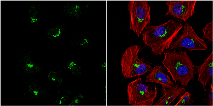
VBP1 antibody detects VBP1 protein at cytoplasm by immunofluorescent analysis. Sample: HeLa cells were fixed in 4% paraformaldehyde at RT for 15 min. Green: VBP1 protein stained by VBP1 antibody (GTX114875) diluted at 1:1000. Red: phalloidin, a cytoskeleton marker, stained by phalloidin (invitrogen, A12380) diluted at 1:200. Blue: Hoechst 33342 staining.
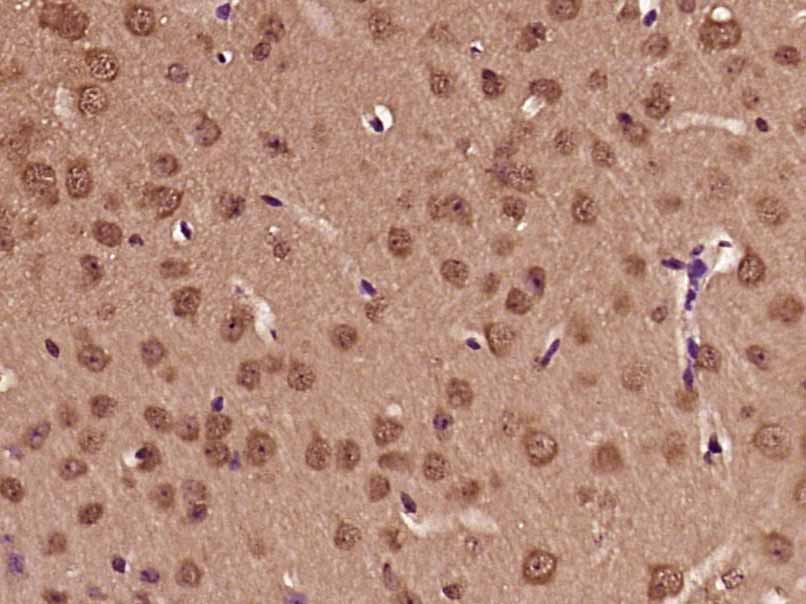
 A: mouse brain 12% SDS PAGE GTX114875 diluted at 1:1000")
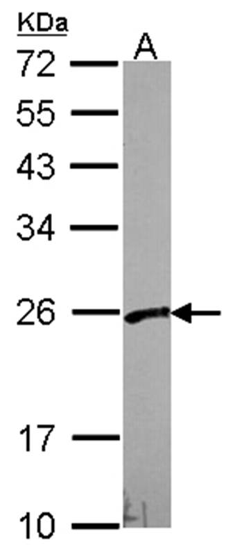
 A: A549 12% SDS PAGE GTX114875 diluted at 1:1000")
VBP1 antibody detects VBP1 protein at cytoplasm by immunofluorescent analysis. Sample: HeLa cells were fixed in 4% paraformaldehyde at RT for 15 min. Green: VBP1 protein stained by VBP1 antibody (GTX114875) diluted at 1:1000. Red: phalloidin, a cytoskeleton marker, stained by phalloidin (invitrogen, A12380) diluted at 1:200. Blue: Hoechst 33342 staining.
PFDN3 antibody
GTX114875
ApplicationsImmunoFluorescence, Western Blot, ImmunoCytoChemistry
Product group Antibodies
ReactivityHuman, Mouse
TargetVBP1
Overview
- SupplierGeneTex
- Product NamePFDN3 antibody
- Delivery Days Customer9
- Application Supplier NoteWB: 1:500-1:3000. ICC/IF: 1:100-1:1000. *Optimal dilutions/concentrations should be determined by the researcher.Not tested in other applications.
- ApplicationsImmunoFluorescence, Western Blot, ImmunoCytoChemistry
- CertificationResearch Use Only
- ClonalityPolyclonal
- Concentration1 mg/ml
- ConjugateUnconjugated
- Gene ID7411
- Target nameVBP1
- Target descriptionVHL binding protein 1
- Target synonymsHIBBJ46, PFD3, PFDN3, VBP-1, prefoldin subunit 3, von Hippel-Lindau binding protein 1
- HostRabbit
- IsotypeIgG
- Protein IDP61758
- Protein NamePrefoldin subunit 3
- Scientific DescriptionThe protein encoded by this gene interacts with the Von Hippel-Lindau protein to form an intracellular complex. Because it functions as a chaperone protein, it is suspected that it may play a role in the transport of the Von Hippel-Lindau protein from the perinuclear granules to the nucleus or cytoplasm. [provided by RefSeq]
- ReactivityHuman, Mouse
- Storage Instruction-20°C or -80°C,2°C to 8°C
- UNSPSC41116161
Datasheet
Related products
Product group Antibodies
VBP1 AntibodyCSB-PA025808GA01HU
ApplicationsImmunoFluorescence, Western Blot, ELISA, ImmunoHistoChemistry
ReactivityHuman, Mouse, Rat
TargetVBP1
- SizePrice

Product group Antibodies
ApplicationsWestern Blot
ReactivityHuman, Mouse
TargetVBP1
- SizePrice

Product group Antibodies
Anti-VBP1 AntibodyHPA023230
ApplicationsWestern Blot, ImmunoCytoChemistry, ImmunoHistoChemistry
ReactivityHuman
TargetVBP1
- SizePrice
Product group Antibodies

VBP1 AntibodyLS-C410524
ApplicationsWestern Blot
ReactivityHuman, Mouse
TargetVBP1
- SizePrice
Product group Antibodies
PFDN3 Polyclonal AntibodyBS-12628R
ApplicationsImmunoFluorescence, Western Blot, ELISA, ImmunoCytoChemistry, ImmunoHistoChemistry, ImmunoHistoChemistry Frozen, ImmunoHistoChemistry Paraffin
ReactivityHuman, Mouse
TargetVBP1
- SizePrice
Product group Antibodies
Anti-VBP1 Antibody144-08990
ApplicationsWestern Blot
ReactivityHuman, Mouse
TargetVBP1
- SizePrice