

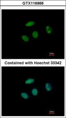
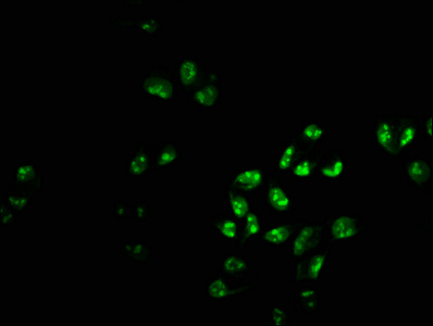
Immunofluorescence analysis of paraformaldehyde-fixed MCF-7, using PHF6(GTX116966) antibody at 1:500 dilution.
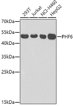
![MCF-7 whole cell and nuclear extracts (30 μg) were separated by 10% SDS-PAGE, and the membrane was blotted with PHF6 antibody [N2C2], Internal (GTX116966) diluted at 1:1000. The HRP-conjugated anti-rabbit IgG antibody (GTX213110-01) was used to detect the primary antibody.](https://www.genetex.com/upload/website/prouct_img/normal/GTX116966/GTX116966_40436_20210806_WB_Fraction_w_23060519_313.webp "MCF-7 whole cell and nuclear extracts (30 μg) were separated by 10% SDS-PAGE, and the membrane was blotted with PHF6 antibody [N2C2], Internal (GTX116966) diluted at 1:1000. The HRP-conjugated anti-rabbit IgG antibody (GTX213110-01) was used to detect the primary antibody.")
Immunofluorescence analysis of paraformaldehyde-fixed MCF-7, using PHF6(GTX116966) antibody at 1:500 dilution.
PHF6 antibody [N2C2], Internal
GTX116966
ApplicationsImmunoFluorescence, Western Blot, ImmunoCytoChemistry
Product group Antibodies
ReactivityHuman
TargetPHF6
Overview
- SupplierGeneTex
- Product NamePHF6 antibody [N2C2], Internal
- Delivery Days Customer9
- Application Supplier NoteWB: 1:500-1:3000. ICC/IF: 1:100-1:1000. *Optimal dilutions/concentrations should be determined by the researcher.Not tested in other applications.
- ApplicationsImmunoFluorescence, Western Blot, ImmunoCytoChemistry
- CertificationResearch Use Only
- ClonalityPolyclonal
- Concentration1 mg/ml
- ConjugateUnconjugated
- Gene ID84295
- Target namePHF6
- Target descriptionPHD finger protein 6
- Target synonymsBFLS, BORJ, CENP-31, PHD finger protein 6, PHD-like zinc finger protein, centromere protein 31
- HostRabbit
- IsotypeIgG
- Protein IDQ8IWS0
- Protein NamePHD finger protein 6
- Scientific DescriptionThis gene is a member of the plant homeodomain (PHD)-like finger (PHF) family. It encodes a protein with two PHD-type zinc finger domains, indicating a potential role in transcriptional regulation, that localizes to the nucleolus. Mutations affecting the coding region of this gene or the splicing of the transcript have been associated with Borjeson-Forssman-Lehmann syndrome (BFLS), a disorder characterized by mental retardation, epilepsy, hypogonadism, hypometabolism, obesity, swelling of subcutaneous tissue of the face, narrow palpebral fissures, and large ears. Alternate transcriptional splice variants, encoding different isoforms, have been characterized. [provided by RefSeq]
- ReactivityHuman
- Storage Instruction-20°C or -80°C,2°C to 8°C
- UNSPSC41116161
Datasheet
Related products
Product group Antibodies
PHF6 AntibodyCSB-PA017917LA01HU
ApplicationsImmunoFluorescence, ImmunoPrecipitation, Western Blot, ELISA, ImmunoHistoChemistry
ReactivityHuman
TargetPHF6
- SizePrice

Product group Antibodies
Anti-PHF6 Antibody Picoband(r)A03065-1-CARRIER-FREE
ApplicationsFlow Cytometry, ImmunoFluorescence, Western Blot, ELISA, ImmunoCytoChemistry, ImmunoHistoChemistry
ReactivityHuman, Monkey, Mouse, Rat
TargetPHF6
- SizePrice
Product group Antibodies
Anti-PHF6 AntibodyA32043
ApplicationsWestern Blot, ImmunoHistoChemistry
ReactivityHuman, Mouse
- SizePrice
Product group Antibodies

PHF6 AntibodyLS-C831742
ApplicationsELISA, ImmunoHistoChemistry
ReactivityHuman, Mouse
TargetPHF6
- SizePrice

Product group Antibodies
Anti-PHF6 AntibodyHPA001023
ApplicationsWestern Blot, ImmunoCytoChemistry, ImmunoHistoChemistry
ReactivityHuman
TargetPHF6
- SizePrice
Product group Antibodies
Phf6 Polyclonal AntibodyCAC08722
ApplicationsImmunoFluorescence, ImmunoPrecipitation, Western Blot, ELISA, ImmunoHistoChemistry
TargetPHF6
- SizePrice
Product group Antibodies
PHF6 antibodyGTX33408
ApplicationsWestern Blot
ReactivityHuman
TargetPHF6
- SizePrice
Product group Antibodies
Anti-PHF6 (Center) Antibody102-24995
ApplicationsWestern Blot
TargetPHF6
- SizePrice