

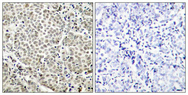
Immunohistochemical analysis of paraffin-embedded human breast carcinoma tissue using NFkB-p65(Phospho-Ser529) Antibody(left) or the same antibody preincubated with blocking peptide(right).
 Antibody.")
, using NFkappaB-p65 (Phospho-Ser529) Antibody.")
Antibody.")
Immunohistochemical analysis of paraffin-embedded human breast carcinoma tissue using NFkB-p65(Phospho-Ser529) Antibody(left) or the same antibody preincubated with blocking peptide(right).
Phospho-RELA (Ser529) Antibody
CSB-PA794822
ApplicationsImmunoFluorescence, Western Blot, ELISA, ImmunoHistoChemistry
Product group Antibodies
ReactivityHuman, Mouse, Rat
TargetRELA
Overview
- SupplierCusabio
- Product NamePhospho-RELA (Ser529) Antibody
- Delivery Days Customer20
- ApplicationsImmunoFluorescence, Western Blot, ELISA, ImmunoHistoChemistry
- CertificationResearch Use Only
- ClonalityPolyclonal
- ConjugateUnconjugated
- Gene ID5970
- Target nameRELA
- Target descriptionRELA proto-oncogene, NF-kB subunit
- Target synonymsCMCU; NF-kappa-B p65delta3; NF-kappa-B transcription factor p65; NFKB3; nuclear factor NF-kappa-B p65 subunit; nuclear factor of kappa light polypeptide gene enhancer in B-cells 3; p65; transcription factor p65; v-rel avian reticuloendotheliosis viral oncogene homolog A
- HostRabbit
- IsotypeIgG
- Protein IDQ04206
- Protein NameTranscription factor p65
- Scientific DescriptionNF-kappa-B is a pleiotropic transcription factor which is present in almost all cell types and is involved in many biological processed such as inflammation, immunity, differentiation, cell growth, tumorigenesis and apoptosis. NF-kappa-B is a homo- or heterodimeric complex formed by the Rel-like domain-containing proteins RELA/p65, RELB, NFKB1/p105, NFKB1/p50, REL and NFKB2/p52 and the heterodimeric p65-p50 complex appears to be most abundant one. The dimers bind at kappa-B sites in the DNA of their target genes and the individual dimers have distinct preferences for different kappa-B sites that they can bind with distinguishable affinity and specificity. Different dimer combinations act as transcriptional activators or repressors, respectively. NF-kappa-B is controlled by various mechanisms of post-translational modification and subcellular compartmentalization as well as by interactions with other cofactors or corepressors. NF-kappa-B complexes are held in the cytoplasm in an inactive state complexed with members of the NF-kappa-B inhibitor (I-kappa-B) family. In a conventional activation pathway, I-kappa-B is phosphorylated by I-kappa-B kinases (IKKs) in response to different activators, subsequently degraded thus liberating the active NF-kappa-B complex which translocates to the nucleus. NF-kappa-B heterodimeric p65-p50 and p65-c-Rel complexes are transcriptional activators. The NF-kappa-B p65-p65 complex appears to be involved in invasin-mediated activation of IL-8 expression. The inhibitory effect of I-kappa-B upon NF-kappa-B the cytoplasm is exerted primarily through the interaction with p65. p65 shows a weak DNA-binding site which could contribute directly to DNA binding in the NF-kappa-B complex Xu C, et al (2005) Oncogene:24(28): 4486-95. McNulty SE, et al. (2004) Pigment Cell Res Apr; 17(2): 173-80. Madrid LV,et al. (2001) J Biol Chem: 276(22): 18934-40. Wang D, et al. (2000) J Biol Chem : 275(42): 32592-7.
- ReactivityHuman, Mouse, Rat
- Storage Instruction-20°C or -80°C
- UNSPSC41116161
Related products
Product group Antibodies
References
NFKB p65 Polyclonal AntibodyBS-0465R
ApplicationsFlow Cytometry, ImmunoFluorescence, Western Blot, ELISA, ImmunoCytoChemistry, ImmunoHistoChemistry, ImmunoHistoChemistry Frozen, ImmunoHistoChemistry Paraffin
TargetRELA
- SizePrice
Product group Antibodies
TargetRELA
- SizePrice
Product group Antibodies
Rela Polyclonal AntibodyCAC07031
ApplicationsImmunoFluorescence, Western Blot, ChIP Chromatin ImmunoPrecipitation, ELISA, ImmunoHistoChemistry
TargetRELA
- SizePrice
Product group Antibodies
ApplicationsImmunoPrecipitation
TargetRELA
- SizePrice
Product group Antibodies

NFKBp65 AntibodyABX030106
ApplicationsImmunoFluorescence, Western Blot, ELISA, ImmunoCytoChemistry
- SizePrice
Product group Antibodies
anti-NF-kB (p65), pAbAG-25T-0004
ApplicationsImmunoPrecipitation, Western Blot, ImmunoCytoChemistry
TargetRELA
- SizePrice

Product group Antibodies
Anti-NF-kB p65/RELA Antibody Picoband(r)A00284-1-CARRIER-FREE
ApplicationsFlow Cytometry, ImmunoFluorescence, Western Blot, ELISA, ImmunoCytoChemistry
TargetRELA
- SizePrice
Product group Antibodies
ApplicationsWestern Blot, ELISA, ImmunoHistoChemistry
- SizePrice
Product group Antibodies
Anti-RELA Antibody144-02547
ApplicationsImmunoFluorescence, Western Blot, ImmunoHistoChemistry
TargetRELA
- SizePrice