


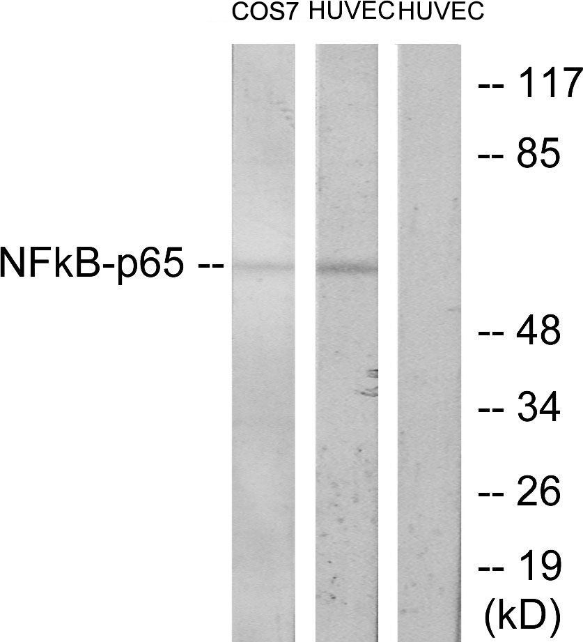
Figure 1. Western blot analysis of NF-kB p65 using anti-NF-kB p65 antibody (A00284-1). Electrophoresis was performed on a 5-20% SDS-PAGE gel at 70V (Stacking gel) / 90V (Resolving gel) for 2-3 hours. The sample well of each lane was loaded with 30 ug of sample under reducing conditions. Lane 1: human Hela whole cell lysates, Lane 2: human Raji whole cell lysates, Lane 3: human HepG2 whole cell lysates, Lane 4: human K562 whole cell lysates, Lane 5: rat PC-12 whole cell lysates, Lane 6: rat RH35 whole cell lysates, Lane 7: mouse RAW264.7 whole cell lysates, Lane 8: mouse HEPA1/6 whole cell lysates. After electrophoresis, proteins were transferred to a nitrocellulose membrane at 150 mA for 50-90 minutes. Blocked the membrane with 5% non-fat milk/TBS for 1.5 hour at RT. The membrane was incubated with rabbit anti-NF-kB p65 antigen affinity purified polyclonal antibody (Catalog # A00284-1) at 0.5 microg/mL overnight at 4°C, then washed with TBS-0.1%Tween 3 times with 5 minutes each and probed with a goat anti-rabbit IgG-HRP secondary antibody at a dilution of 1:5000 for 1.5 hour at RT. The signal is developed using an Enhanced Chemiluminescent detection (ECL) kit (Catalog # EK1002) with Tanon 5200 system. A specific band was detected for NF-kB p65 at approximately 65-70 kDa. The expected band size for NF-kB p65 is at 60 kDa.
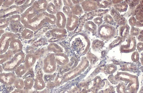
. NF-kB p65 was detected in an immunocytochemical section of U2OS cells. Enzyme antigen retrieval was performed using IHC enzyme antigen retrieval reagent (AR0022) for 15 mins. The cells were blocked with 10% goat serum. And then incubated with 5 microg/mL rabbit anti-NF-kB p65 Antibody (A00284-1) overnight at 4°C. DyLight®488 Conjugated Goat Anti-Rabbit IgG (BA1127) was used as secondary antibody at 1:500 dilution and incubated for 30 minutes at 37°C. The section was counterstained with DAPI. Visualize using a fluorescence microscope and filter sets appropriate for the label used.")
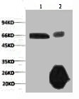
. Lane 1: HepG2 whole cell lysates (30ug), Lane 2: Rabbit control IgG instead of anti-NF-kB p65 antibody in HepG2 whole cell lysate, Lane 3: anti-NF-kB p65 antibody (2microg) + HepG2 whole cell lysate (500microg). After electrophoresis, proteins were transferred to a membrane. Then the membrane was incubated with rabbit anti-NF-kB p65 antigen affinity purified polyclonal antibody (A00284-1) at a dilution of 0.5 microg/mL and probed with a goat anti-rabbit IgG-HRP secondary antibody (Catalog # BA1054). The signal is developed using ECL Plus Western Blotting Substrate (Catalog # AR1197). A specific band was detected for NF-kB p65 at approximately 65-70 kDa. The expected band size for NF-kB p65 is at 60 kDa.")
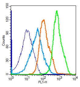
. Overlay histogram showing Hela cells stained with A00284-1 (Blue line). To facilitate intracellular staining, cells were fixed with 4% paraformaldehyde and permeabilized with permeabilization buffer. The cells were blocked with 10% normal goat serum. And then incubated with rabbit anti-NF-kB p65 Antibody (A00284-1, 1 microg/1x106 cells) for 30 min at 20°C. DyLight®488 conjugated goat anti-rabbit IgG (BA1127, 5-10 microg/1x106 cells) was used as secondary antibody for 30 minutes at 20°C. Isotype control antibody (Green line) was rabbit IgG (1 microg/1x106) used under the same conditions. Unlabelled sample without incubation with primary antibody and secondary antibody (Red line) was used as a blank control.")
Figure 1. Western blot analysis of NF-kB p65 using anti-NF-kB p65 antibody (A00284-1). Electrophoresis was performed on a 5-20% SDS-PAGE gel at 70V (Stacking gel) / 90V (Resolving gel) for 2-3 hours. The sample well of each lane was loaded with 30 ug of sample under reducing conditions. Lane 1: human Hela whole cell lysates, Lane 2: human Raji whole cell lysates, Lane 3: human HepG2 whole cell lysates, Lane 4: human K562 whole cell lysates, Lane 5: rat PC-12 whole cell lysates, Lane 6: rat RH35 whole cell lysates, Lane 7: mouse RAW264.7 whole cell lysates, Lane 8: mouse HEPA1/6 whole cell lysates. After electrophoresis, proteins were transferred to a nitrocellulose membrane at 150 mA for 50-90 minutes. Blocked the membrane with 5% non-fat milk/TBS for 1.5 hour at RT. The membrane was incubated with rabbit anti-NF-kB p65 antigen affinity purified polyclonal antibody (Catalog # A00284-1) at 0.5 microg/mL overnight at 4°C, then washed with TBS-0.1%Tween 3 times with 5 minutes each and probed with a goat anti-rabbit IgG-HRP secondary antibody at a dilution of 1:5000 for 1.5 hour at RT. The signal is developed using an Enhanced Chemiluminescent detection (ECL) kit (Catalog # EK1002) with Tanon 5200 system. A specific band was detected for NF-kB p65 at approximately 65-70 kDa. The expected band size for NF-kB p65 is at 60 kDa.
Anti-NF-kB p65/RELA Antibody Picoband(r)
A00284-1-CARRIER-FREE
ApplicationsFlow Cytometry, ImmunoFluorescence, Western Blot, ELISA, ImmunoCytoChemistry
Product group Antibodies
ReactivityHuman, Mouse, Rat
TargetRELA
Overview
- SupplierBoster Bio
- Product NameAnti-NF-kB p65/RELA Antibody Picoband(r)
- Delivery Days Customer9
- ApplicationsFlow Cytometry, ImmunoFluorescence, Western Blot, ELISA, ImmunoCytoChemistry
- CertificationResearch Use Only
- ClonalityPolyclonal
- Concentration500 ug/ml
- Gene ID5970
- Target nameRELA
- Target descriptionRELA proto-oncogene, NF-kB subunit
- Target synonymsAIF3BL3, CMCU, NFKB3, p65, transcription factor p65, NF-kappa-B p65delta3, NF-kappa-B transcription factor p65, nuclear factor NF-kappa-B p65 subunit, nuclear factor of kappa light polypeptide gene enhancer in B-cells 3, v-rel avian reticuloendotheliosis viral oncogene homolog A
- HostRabbit
- IsotypeIgG
- Protein IDQ04206
- Protein NameTranscription factor p65
- Scientific DescriptionBoster Bio Anti-NF-kB p65/RELA Antibody Picoband® catalog # A00284-1. Tested in ELISA, Flow Cytometry, IF, ICC, WB applications. This antibody reacts with Human, Mouse, Rat. The brand Picoband indicates this is a premium antibody that guarantees superior quality, high affinity, and strong signals with minimal background in Western blot applications. Only our best-performing antibodies are designated as Picoband, ensuring unmatched performance.
- ReactivityHuman, Mouse, Rat
- Storage Instruction-20°C,2°C to 8°C
- UNSPSC12352203
Related products
Product group Antibodies
ApplicationsWestern Blot, ELISA, ImmunoHistoChemistry
ReactivityHuman, Mouse, Rat
- SizePrice
Product group Antibodies
anti-NF-kB (p65), pAbAG-25T-0004
ApplicationsImmunoPrecipitation, Western Blot, ImmunoCytoChemistry
ReactivityHuman, Mouse
TargetRELA
- SizePrice
Product group Antibodies
Anti-RELA Antibody144-02547
ApplicationsImmunoFluorescence, Western Blot, ImmunoHistoChemistry
ReactivityHuman, Rat
TargetRELA
- SizePrice
Product group Antibodies

NFKBp65 AntibodyABX030106
ApplicationsImmunoFluorescence, Western Blot, ELISA, ImmunoCytoChemistry
- SizePrice
Product group Antibodies

RELA / NFKB p65 AntibodyLS-C831476
ApplicationsImmunoHistoChemistry
ReactivityHuman
TargetRELA
- SizePrice
Product group Antibodies
References
NFKB p65 Polyclonal AntibodyBS-0465R
ApplicationsFlow Cytometry, ImmunoFluorescence, Western Blot, ELISA, ImmunoCytoChemistry, ImmunoHistoChemistry, ImmunoHistoChemistry Frozen, ImmunoHistoChemistry Paraffin
ReactivityBovine, Canine, Chicken, Equine, Human, Mouse, Porcine, Rabbit, Rat, Zebra Fish
TargetRELA
- SizePrice
Product group Antibodies
RELA Monoclonal AntibodyCSB-MA000209
ApplicationsImmunoFluorescence, ImmunoPrecipitation, Western Blot, ELISA, ImmunoHistoChemistry
ReactivityHuman, Mouse, Rat
TargetRELA
- SizePrice
Product group Antibodies
Rela Polyclonal AntibodyCAC07031
ApplicationsImmunoFluorescence, Western Blot, ChIP Chromatin ImmunoPrecipitation, ELISA, ImmunoHistoChemistry
ReactivityMouse
TargetRELA
- SizePrice
Product group Antibodies
NFkB p65 antibodyGTX102090
ApplicationsImmunoFluorescence, ImmunoPrecipitation, Western Blot, ChIP Chromatin ImmunoPrecipitation, ImmunoCytoChemistry, ImmunoHistoChemistry, ImmunoHistoChemistry Frozen, ImmunoHistoChemistry Paraffin
ReactivityHuman, Mouse, Rat
TargetRELA
- SizePrice