

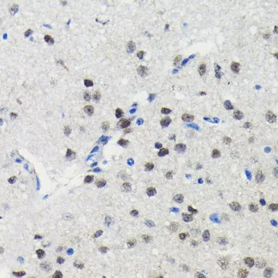
IHC-P analysis of mouse brain tissue section using GTX00954 PIM1 antibody [GT1192]. Dilution : 1:100
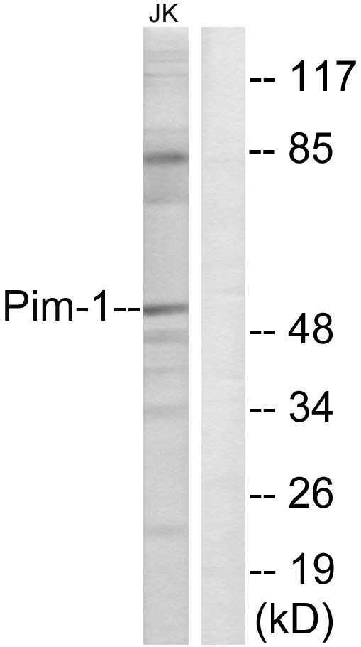
![Various whole cell extracts (30 μg) were separated by 10% SDS-PAGE, and the membrane was blotted with PIM1 antibody [GT1192] (GTX00954) diluted at 1:1000. The HRP-conjugated anti-rabbit IgG antibody (GTX213110-01) was used to detect the primary antibody.](https://www.genetex.com/upload/website/prouct_img/normal/GTX00954/GTX00954_4000000175_20200410_WB_w_23053121_688.webp "Various whole cell extracts (30 μg) were separated by 10% SDS-PAGE, and the membrane was blotted with PIM1 antibody [GT1192] (GTX00954) diluted at 1:1000. The HRP-conjugated anti-rabbit IgG antibody (GTX213110-01) was used to detect the primary antibody.")
![WB analysis of 22Rv1 whole cell lysate using GTX00954 PIM1 antibody [GT1192]. Dilution : 1:1000 Loading : 25 μg](https://www.genetex.com/upload/website/prouct_img/normal/GTX00954/GTX00954_20200508_WB_w_23053121_371.webp "WB analysis of 22Rv1 whole cell lysate using GTX00954 PIM1 antibody [GT1192]. Dilution : 1:1000 Loading : 25 μg")
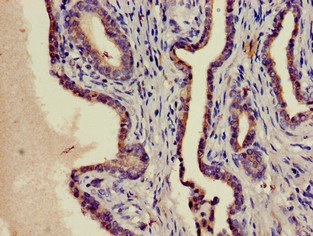
![IHC-P analysis of rat ovary tissue section using GTX00954 PIM1 antibody [GT1192]. Dilution : 1:100](https://www.genetex.com/upload/website/prouct_img/normal/GTX00954/GTX00954_20200508_IHC-P_2_w_23053121_118.webp "IHC-P analysis of rat ovary tissue section using GTX00954 PIM1 antibody [GT1192]. Dilution : 1:100")
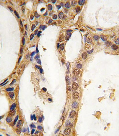
![IHC-P analysis of human colon tissue section using GTX00954 PIM1 antibody [GT1192]. Dilution : 1:100](https://www.genetex.com/upload/website/prouct_img/normal/GTX00954/GTX00954_20200508_IHC-P_1_w_23053121_102.webp "IHC-P analysis of human colon tissue section using GTX00954 PIM1 antibody [GT1192]. Dilution : 1:100")
IHC-P analysis of mouse brain tissue section using GTX00954 PIM1 antibody [GT1192]. Dilution : 1:100
PIM1 antibody [GT1192]
GTX00954
ApplicationsWestern Blot, ImmunoHistoChemistry, ImmunoHistoChemistry Paraffin
Product group Antibodies
ReactivityHuman, Mouse, Rat
TargetPIM1
Overview
- SupplierGeneTex
- Product NamePIM1 antibody [GT1192]
- Delivery Days Customer9
- Application Supplier NoteWB: 1:500 - 1:2000. IHC-P: 1:50 - 1:200. *Optimal dilutions/concentrations should be determined by the researcher.Not tested in other applications.
- ApplicationsWestern Blot, ImmunoHistoChemistry, ImmunoHistoChemistry Paraffin
- CertificationResearch Use Only
- ClonalityMonoclonal
- Clone IDGT1192
- ConjugateUnconjugated
- Gene ID5292
- Target namePIM1
- Target descriptionPim-1 proto-oncogene, serine/threonine kinase
- Target synonymsPIM, serine/threonine-protein kinase pim-1, Oncogene PIM1, pim-1 oncogene (proviral integration site 1), proto-oncogene serine/threonine-protein kinase pim-1
- HostRabbit
- IsotypeIgG
- Protein IDP11309
- Protein NameSerine/threonine-protein kinase pim-1
- Scientific DescriptionThe protein encoded by this gene belongs to the Ser/Thr protein kinase family, and PIM subfamily. This gene is expressed primarily in B-lymphoid and myeloid cell lines, and is overexpressed in hematopoietic malignancies and in prostate cancer. It plays a role in signal transduction in blood cells, contributing to both cell proliferation and survival, and thus provides a selective advantage in tumorigenesis. Both the human and orthologous mouse genes have been reported to encode two isoforms (with preferential cellular localization) resulting from the use of alternative in-frame translation initiation codons, the upstream non-AUG (CUG) and downstream AUG codons (PMIDs:16186805, 1825810).[provided by RefSeq, Aug 2011]
- ReactivityHuman, Mouse, Rat
- Storage Instruction-20°C or -80°C,2°C to 8°C
- UNSPSC41116161
Datasheet
Related products
Product group Antibodies
Anti-Pim-1 AntibodyA97345
ApplicationsWestern Blot, ELISA
ReactivityHuman, Mouse, Rat
- SizePrice
Product group Antibodies

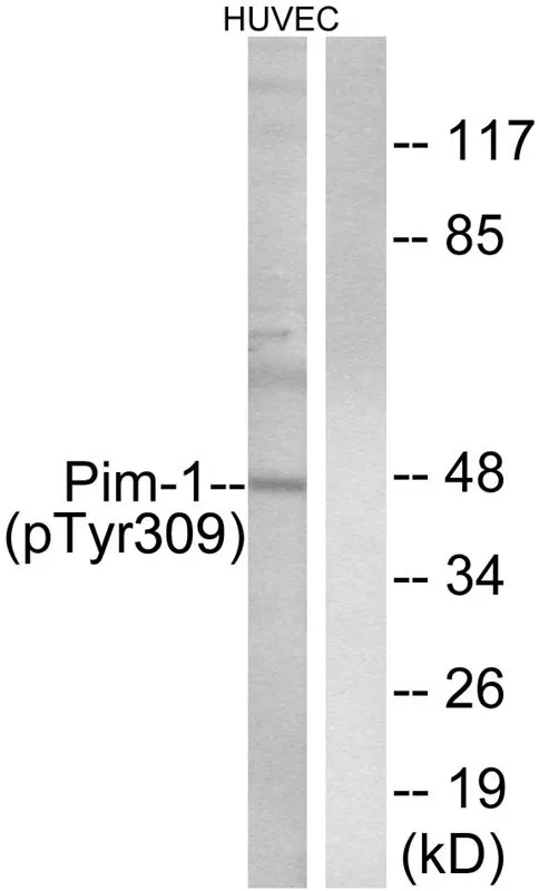
Pim-1 (Phospho-Tyr309) AntibodyABX012463
ApplicationsWestern Blot, ELISA
- SizePrice
Product group Antibodies
Anti-PIM1 Antibody144-60012
ApplicationsWestern Blot
ReactivityHuman, Rat
TargetPIM1
- SizePrice
Product group Antibodies
PIM1 Recombinant AntibodyBSM-61220R
ApplicationsWestern Blot
TargetPIM1
- SizePrice
Product group Antibodies
PIM1 Polyclonal AntibodyCAC13255
ApplicationsImmunoFluorescence, ELISA, ImmunoHistoChemistry
TargetPIM1
- SizePrice
Product group Antibodies
PIM1 AntibodyCSB-PA02959A0RB
ApplicationsImmunoFluorescence, ELISA, ImmunoHistoChemistry
ReactivityHuman
TargetPIM1
- SizePrice
Product group Antibodies

PIM1 / Pim-1 AntibodyLS-C401361
ApplicationsELISA, ImmunoHistoChemistry
ReactivityHuman, Mouse, Rat
TargetPIM1
- SizePrice
Product group Antibodies
PIM1 antibodyGTX81239
ApplicationsWestern Blot, ImmunoHistoChemistry, ImmunoHistoChemistry Paraffin
ReactivityHuman
TargetPIM1
- SizePrice
Product group Antibodies
PIM1 (phospho Tyr309) antibodyGTX55311
ApplicationsWestern Blot
ReactivityHuman
TargetPIM1
- SizePrice