

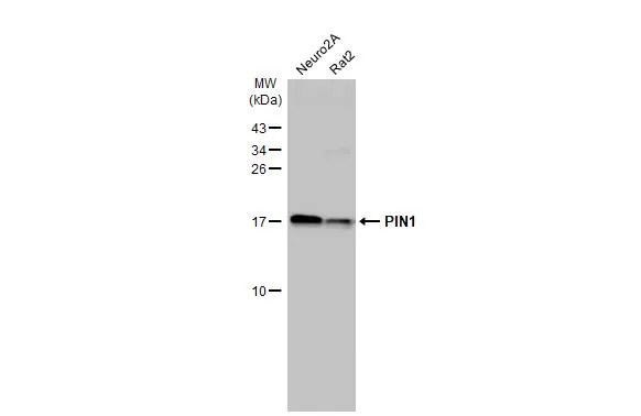
Various whole cell extracts (30 μg) were separated by 15% SDS-PAGE, and the membrane was blotted with PIN1 antibody [HL2480] (GTX638829) diluted at 1:1000. The HRP-conjugated anti-rabbit IgG antibody (GTX213110-01) was used to detect the primary antibody.
![PIN1 antibody [HL2480] detects PIN1 protein at cytoplasm and nucleus by immunohistochemical analysis. Sample: Paraffin-embedded mouse intestine. PIN1 stained by PIN1 antibody [HL2480] (GTX638829) diluted at 1:100. Antigen Retrieval: Citrate buffer, pH 6.0, 15 min](https://www.genetex.com/upload/website/prouct_img/normal/GTX638829/GTX638829_T-45096_20230721_IHC-P_M_23073119_662.webp "PIN1 antibody [HL2480] detects PIN1 protein at cytoplasm and nucleus by immunohistochemical analysis. Sample: Paraffin-embedded mouse intestine. PIN1 stained by PIN1 antibody [HL2480] (GTX638829) diluted at 1:100. Antigen Retrieval: Citrate buffer, pH 6.0, 15 min")
![PIN1 antibody [HL2480] detects PIN1 protein by immunohistochemical analysis. Sample: Paraffin-embedded rat tissues. PIN1 stained by PIN1 antibody [HL2480] (GTX638829) diluted at 1:100. Antigen Retrieval: Citrate buffer, pH 6.0, 15 min](https://www.genetex.com/upload/website/prouct_img/normal/GTX638829/GTX638829_T-45096_20230721_IHC-P_multiple_R_23073119_894.webp "PIN1 antibody [HL2480] detects PIN1 protein by immunohistochemical analysis. Sample: Paraffin-embedded rat tissues. PIN1 stained by PIN1 antibody [HL2480] (GTX638829) diluted at 1:100. Antigen Retrieval: Citrate buffer, pH 6.0, 15 min")
![PIN1 antibody [HL2480] detects PIN1 protein at cytoplasm and nucleus by immunohistochemical analysis. Sample: Paraffin-embedded human breast carcinoma. PIN1 stained by PIN1 antibody [HL2480] (GTX638829) diluted at 1:100. Antigen Retrieval: Citrate buffer, pH 6.0, 15 min](https://www.genetex.com/upload/website/prouct_img/normal/GTX638829/GTX638829_T-45096_20230721_IHC-P_23073119_918.webp "PIN1 antibody [HL2480] detects PIN1 protein at cytoplasm and nucleus by immunohistochemical analysis. Sample: Paraffin-embedded human breast carcinoma. PIN1 stained by PIN1 antibody [HL2480] (GTX638829) diluted at 1:100. Antigen Retrieval: Citrate buffer, pH 6.0, 15 min")
![Whole zebrafish extract (30 μg) was separated by 15% SDS-PAGE, and the membrane was blotted with PIN1 antibody [HL2480] (GTX638829) diluted at 1:1000. The HRP-conjugated anti-rabbit IgG antibody (GTX213110-01) was used to detect the primary antibody.](https://www.genetex.com/upload/website/prouct_img/normal/GTX638829/GTX638829_45201_20231027_WB_Z_23103019_315.webp "Whole zebrafish extract (30 μg) was separated by 15% SDS-PAGE, and the membrane was blotted with PIN1 antibody [HL2480] (GTX638829) diluted at 1:1000. The HRP-conjugated anti-rabbit IgG antibody (GTX213110-01) was used to detect the primary antibody.")
![Various whole cell extracts (30 μg) were separated by 15% SDS-PAGE, and the membrane was blotted with PIN1 antibody [HL2480] (GTX638829) diluted at 1:1000. The HRP-conjugated anti-rabbit IgG antibody (GTX213110-01) was used to detect the primary antibody, and the signal was developed with Trident ECL plus-Enhanced.](https://www.genetex.com/upload/website/prouct_img/normal/GTX638829/GTX638829_45201_20231027_WB_23103019_916.webp "Various whole cell extracts (30 μg) were separated by 15% SDS-PAGE, and the membrane was blotted with PIN1 antibody [HL2480] (GTX638829) diluted at 1:1000. The HRP-conjugated anti-rabbit IgG antibody (GTX213110-01) was used to detect the primary antibody, and the signal was developed with Trident ECL plus-Enhanced.")
![PIN1 antibody [HL2480] detects PIN1 protein at cytoplasm and nucleus by immunofluorescent analysis. Sample: NIH-3T3 cells were fixed in 4% paraformaldehyde at RT for 15 min. Green: PIN1 stained by PIN1 antibody [HL2480] (GTX638829) diluted at 1:500. Red: alpha Tubulin, a cytoskeleton marker, stained by alpha Tubulin antibody [GT114] (GTX628802) diluted at 1:1000. Blue: Fluoroshield with DAPI (GTX30920).](https://www.genetex.com/upload/website/prouct_img/normal/GTX638829/GTX638829_T-45159_20240202_ICC_IF_24021917_571.webp "PIN1 antibody [HL2480] detects PIN1 protein at cytoplasm and nucleus by immunofluorescent analysis. Sample: NIH-3T3 cells were fixed in 4% paraformaldehyde at RT for 15 min. Green: PIN1 stained by PIN1 antibody [HL2480] (GTX638829) diluted at 1:500. Red: alpha Tubulin, a cytoskeleton marker, stained by alpha Tubulin antibody [GT114] (GTX628802) diluted at 1:1000. Blue: Fluoroshield with DAPI (GTX30920).")
Various whole cell extracts (30 μg) were separated by 15% SDS-PAGE, and the membrane was blotted with PIN1 antibody [HL2480] (GTX638829) diluted at 1:1000. The HRP-conjugated anti-rabbit IgG antibody (GTX213110-01) was used to detect the primary antibody.
PIN1 antibody [HL2480]
GTX638829
ApplicationsImmunoFluorescence, Western Blot, ImmunoCytoChemistry, ImmunoHistoChemistry, ImmunoHistoChemistry Paraffin
Product group Antibodies
ReactivityHuman, Mouse, Rat, Zebra Fish
TargetPIN1
Overview
- SupplierGeneTex
- Product NamePIN1 antibody [HL2480]
- Delivery Days Customer9
- Application Supplier NoteWB: 1:500-1:3000. *Optimal dilutions/concentrations should be determined by the researcher.Not tested in other applications.
- ApplicationsImmunoFluorescence, Western Blot, ImmunoCytoChemistry, ImmunoHistoChemistry, ImmunoHistoChemistry Paraffin
- CertificationResearch Use Only
- ClonalityMonoclonal
- Clone IDHL2480
- Concentration1 mg/ml
- ConjugateUnconjugated
- Gene ID5300
- Target namePIN1
- Target descriptionpeptidylprolyl cis/trans isomerase, NIMA-interacting 1
- Target synonymsDOD, UBL5, peptidyl-prolyl cis-trans isomerase NIMA-interacting 1, PPIase Pin1, parvulin PIN1, protein (peptidyl-prolyl cis/trans isomerase) NIMA-interacting 1, protein interacting with never in mitosis A1, rotamase Pin1
- HostRabbit
- IsotypeIgG
- Protein IDQ13526
- Protein NamePeptidyl-prolyl cis-trans isomerase NIMA-interacting 1
- Scientific DescriptionPeptidyl-prolyl cis/trans isomerases (PPIases) catalyze the cis/trans isomerization of peptidyl-prolyl peptide bonds. This gene encodes one of the PPIases, which specifically binds to phosphorylated ser/thr-pro motifs to catalytically regulate the post-phosphorylation conformation of its substrates. The conformational regulation catalyzed by this PPIase has a profound impact on key proteins involved in the regulation of cell growth, genotoxic and other stress responses, the immune response, induction and maintenance of pluripotency, germ cell development, neuronal differentiation, and survival. This enzyme also plays a key role in the pathogenesis of Alzheimers disease and many cancers. Multiple alternatively spliced transcript variants have been found for this gene.[provided by RefSeq, Jun 2011]
- ReactivityHuman, Mouse, Rat, Zebra Fish
- Storage Instruction-20°C or -80°C,2°C to 8°C
- UNSPSC41116161
Datasheet
Related products
Product group Antibodies
PIN1 Polyclonal AntibodyCAC14064
ApplicationsImmunoPrecipitation, ELISA, ImmunoHistoChemistry
TargetPIN1
- SizePrice
Product group Antibodies
Anti-Pin1 AntibodyA84260
ApplicationsWestern Blot, ELISA
ReactivityHuman, Rat
- SizePrice
Product group Antibodies

Goat anti-PIN1EB07468
ApplicationsWestern Blot, ELISA
ReactivityCanine, Human, Mouse, Rat
TargetPIN1
- SizePrice
Product group Antibodies
Phospho-PIN1 (S16) AntibodyCSB-PA030061
ApplicationsWestern Blot, ELISA, ImmunoHistoChemistry
ReactivityHuman, Monkey, Mouse, Rat
TargetPIN1
- SizePrice

Product group Antibodies
Anti-PIN1 AntibodyHPA068650
ApplicationsWestern Blot, ImmunoCytoChemistry
ReactivityHuman
TargetPIN1
- SizePrice
Product group Antibodies

PIN1 Antibody (N-Terminus)LS-C368471
ApplicationsImmunoFluorescence, Western Blot, ImmunoCytoChemistry, ImmunoHistoChemistry, ImmunoHistoChemistry Paraffin
ReactivityBovine, Human, Monkey, Mouse, Porcine, Rat, Zebra Fish
TargetPIN1
- SizePrice

Product group Antibodies
Anti-Pin1 Antibody Picoband(r)PB9316-CARRIER-FREE
ApplicationsFlow Cytometry, Western Blot
ReactivityHuman, Monkey, Mouse, Rat
TargetPIN1
- SizePrice
![Various whole cell extracts (30 μg) were separated by 15% SDS-PAGE, and the membrane was blotted with PIN1 antibody [HL2115] (GTX638087) diluted at 1:1000. The HRP-conjugated anti-rabbit IgG antibody (GTX213110-01) was used to detect the primary antibody, and the signal was developed with Trident ECL plus-Enhanced.](https://www.genetex.com/upload/website/prouct_img/normal/GTX638087/GTX638087_T-44907_20221230_WB_23010400_538.webp)
Product group Antibodies
PIN1 antibody [HL2115]GTX638087
ApplicationsImmunoFluorescence, Western Blot, ImmunoCytoChemistry
ReactivityHuman, Mouse, Rat
TargetPIN1
- SizePrice