

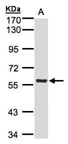
Sample(30 ug whole cell lysate) A:A431(GTX27909) 7.5% SDS PAGE GTX107851 diluted at 1:1000
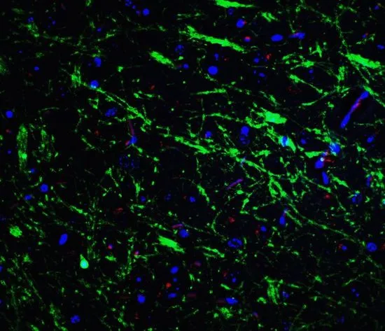
![PINK1 antibody [N3C3] detects PINK1 protein at mitochondria by immunofluorescent analysis. Sample: MCF-7 cells were fixed in ice-cold MeOH for 5 min. Green: PINK1 protein stained by PINK1 antibody [N3C3] (GTX107851) diluted at 1:500. Blue: Hoechst 33342 staining.](https://www.genetex.com/upload/website/prouct_img/normal/GTX107851/GTX107851_40002_IFA_w_23060120_341.webp "PINK1 antibody [N3C3] detects PINK1 protein at mitochondria by immunofluorescent analysis. Sample: MCF-7 cells were fixed in ice-cold MeOH for 5 min. Green: PINK1 protein stained by PINK1 antibody [N3C3] (GTX107851) diluted at 1:500. Blue: Hoechst 33342 staining.")

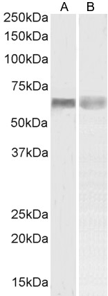
![Untreated (–) and treated (+) PC-3 whole cell extracts (30 μg) were separated by 7.5% SDS-PAGE, and the membrane was blotted with PINK1 antibody [N3C3] (GTX107851) diluted at 1:1000. The HRP-conjugated anti-rabbit IgG antibody (GTX213110-01) was used to detect the primary antibody.](https://www.genetex.com/upload/website/prouct_img/normal/GTX107851/GTX107851_45196_20250523_WB_treatment_CCCP_25052800_931.webp "Untreated (–) and treated (+) PC-3 whole cell extracts (30 μg) were separated by 7.5% SDS-PAGE, and the membrane was blotted with PINK1 antibody [N3C3] (GTX107851) diluted at 1:1000. The HRP-conjugated anti-rabbit IgG antibody (GTX213110-01) was used to detect the primary antibody.")
Sample(30 ug whole cell lysate) A:A431(GTX27909) 7.5% SDS PAGE GTX107851 diluted at 1:1000
PINK1 antibody [N3C3]
GTX107851
ApplicationsImmunoFluorescence, Western Blot, ImmunoCytoChemistry
Product group Antibodies
ReactivityHuman, Mouse, Rat
TargetPINK1
Overview
- SupplierGeneTex
- Product NamePINK1 antibody [N3C3]
- Delivery Days Customer9
- Application Supplier NoteWB: 1:500-1:3000. ICC/IF: 1:100-1:1000. *Optimal dilutions/concentrations should be determined by the researcher.Not tested in other applications.
- ApplicationsImmunoFluorescence, Western Blot, ImmunoCytoChemistry
- CertificationResearch Use Only
- ClonalityPolyclonal
- Concentration1.37 mg/ml
- ConjugateUnconjugated
- Gene ID65018
- Target namePINK1
- Target descriptionPTEN induced kinase 1
- Target synonymsBRPK, PARK6, serine/threonine-protein kinase PINK1, mitochondrial, PTEN induced putative kinase 1, PTEN-induced putative kinase protein 1, protein kinase BRPK
- HostRabbit
- IsotypeIgG
- Protein IDQ9BXM7
- Protein NameSerine/threonine-protein kinase PINK1, mitochondrial
- Scientific DescriptionThis gene encodes a serine/threonine protein kinase that localizes to mitochondria. It is thought to protect cells from stress-induced mitochondrial dysfunction. Mutations in this gene cause one form of autosomal recessive early-onset Parkinson disease. [provided by RefSeq]
- ReactivityHuman, Mouse, Rat
- Storage Instruction-20°C or -80°C,2°C to 8°C
- UNSPSC41116161
Datasheet
Related products
Product group Antibodies
Anti-PINK1 AntibodyA84378
ApplicationsFlow Cytometry, Western Blot, ELISA
ReactivityHuman
- SizePrice
Product group Antibodies
Anti-PINK1 Antibody144-66301
ApplicationsImmunoFluorescence, Western Blot
ReactivityHuman, Mouse, Rat
TargetPINK1
- SizePrice
Product group Antibodies
PINK1 AntibodyCSB-PA447054
ApplicationsELISA
ReactivityHuman, Mouse
TargetPINK1
- SizePrice
Product group Antibodies

PINK1 AntibodyLS-C761174
ApplicationsWestern Blot
ReactivityHuman, Mouse, Rat
TargetPINK1
- SizePrice

Product group Antibodies
Anti-PINK1 Antibody Picoband(r)A00201-2-CARRIER-FREE
ApplicationsFlow Cytometry, ImmunoFluorescence, Western Blot, ELISA, ImmunoCytoChemistry
ReactivityHuman
TargetPINK1
- SizePrice
Product group Antibodies
PINK1 Polyclonal AntibodyBS-22173R
ApplicationsWestern Blot
ReactivityHuman, Mouse, Rat
TargetPINK1
- SizePrice
Product group Antibodies

Goat anti-PINK1EB07940
ApplicationsFlow Cytometry, Western Blot, ELISA
ReactivityHuman, Rat
TargetPINK1
- SizePrice
Product group Antibodies
Pink1 Polyclonal AntibodyCAC10977
ApplicationsELISA, ImmunoHistoChemistry
TargetPINK1
- SizePrice
Product group Antibodies
PINK1 antibodyGTX31581
ApplicationsWestern Blot, ELISA, ImmunoHistoChemistry, ImmunoHistoChemistry Paraffin
ReactivityHuman, Mouse, Rat
TargetPINK1
- SizePrice
![WB analysis of rat brain tissue lysate using GTX04667 PINK1 antibody [S4-15]. Loading : 15 μg Dilution : 1:200](https://www.genetex.com/upload/website/prouct_img/normal/GTX04667/GTX04667_20231214_WB_23121400_650.webp)
Product group Antibodies
PINK1 antibody [S4-15]GTX04667
ApplicationsImmunoFluorescence, Western Blot, ImmunoCytoChemistry
ReactivityHuman, Mouse, Rat
TargetPINK1
- SizePrice