

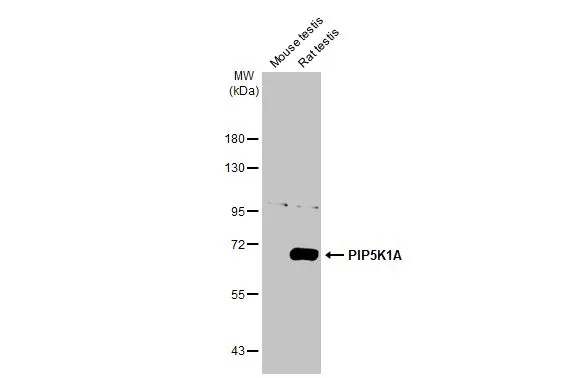
Various tissue extracts (50 μg) were separated by 7.5% SDS-PAGE, and the membrane was blotted with PIP5K1A antibody [HL2011] (GTX637912) diluted at 1:1000. The HRP-conjugated anti-rabbit IgG antibody (GTX213110-01) was used to detect the primary antibody.
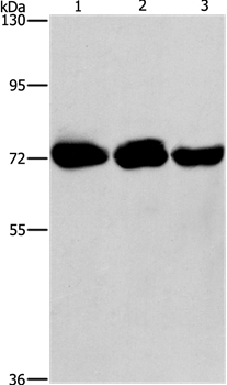
![Whole cell extract (30 μg) was separated by 7.5% SDS-PAGE, and the membrane was blotted with PIP5K1A antibody [HL2011] (GTX637912) diluted at 1:1000. The HRP-conjugated anti-rabbit IgG antibody (GTX213110-01) was used to detect the primary antibody, and the signal was developed with Trident ECL plus-Enhanced.](https://www.genetex.com/upload/website/prouct_img/normal/GTX637912/GTX637912_T-44872_20221209_WB_22121123_362.webp "Whole cell extract (30 μg) was separated by 7.5% SDS-PAGE, and the membrane was blotted with PIP5K1A antibody [HL2011] (GTX637912) diluted at 1:1000. The HRP-conjugated anti-rabbit IgG antibody (GTX213110-01) was used to detect the primary antibody, and the signal was developed with Trident ECL plus-Enhanced.")
![Non-transfected (–) and transfected (+) 293T whole cell extracts were separated by 7.5% SDS-PAGE, and the membrane was blotted with PIP5K1A antibody [HL2011] (GTX637912) diluted at 1:1000. The HRP-conjugated anti-rabbit IgG antibody (GTX213110-01) was used to detect the primary antibody.](https://www.genetex.com/upload/website/prouct_img/normal/GTX637912/GTX637912_T-44872_20221216_WB_multiple_B_22122018_356.webp "Non-transfected (–) and transfected (+) 293T whole cell extracts were separated by 7.5% SDS-PAGE, and the membrane was blotted with PIP5K1A antibody [HL2011] (GTX637912) diluted at 1:1000. The HRP-conjugated anti-rabbit IgG antibody (GTX213110-01) was used to detect the primary antibody.")
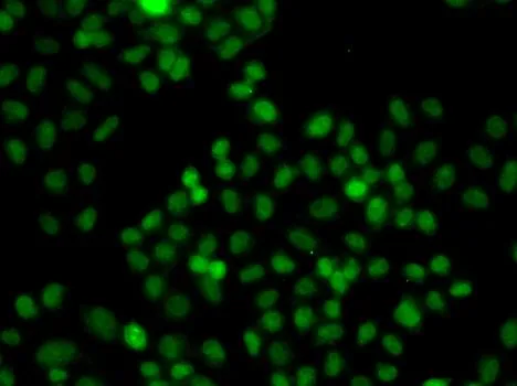
![PIP5K1A antibody [HL2011] detects PIP5K1A protein at cytoplasm and nucleus by immunofluorescent analysis. Sample: HeLa cells were fixed in 4% paraformaldehyde at RT for 15 min. Green: PIP5K1A stained by PIP5K1A antibody [HL2011] (GTX637912) diluted at 1:500. Blue: Fluoroshield with DAPI (GTX30920).](https://www.genetex.com/upload/website/prouct_img/normal/GTX637912/GTX637912_T-44872_20230119_ICC_IF_23021401_792.webp "PIP5K1A antibody [HL2011] detects PIP5K1A protein at cytoplasm and nucleus by immunofluorescent analysis. Sample: HeLa cells were fixed in 4% paraformaldehyde at RT for 15 min. Green: PIP5K1A stained by PIP5K1A antibody [HL2011] (GTX637912) diluted at 1:500. Blue: Fluoroshield with DAPI (GTX30920).")
![Whole cell extract (30 μg) was separated by 7.5% SDS-PAGE, and the membrane was blotted with PIP5K1A antibody [HL2011] (GTX637912) diluted at 1:1000. The HRP-conjugated anti-rabbit IgG antibody (GTX213110-01) was used to detect the primary antibody.](https://www.genetex.com/upload/website/prouct_img/normal/GTX637912/GTX637912_44935_20230616_WB_D_23062019_987.webp "Whole cell extract (30 μg) was separated by 7.5% SDS-PAGE, and the membrane was blotted with PIP5K1A antibody [HL2011] (GTX637912) diluted at 1:1000. The HRP-conjugated anti-rabbit IgG antibody (GTX213110-01) was used to detect the primary antibody.")
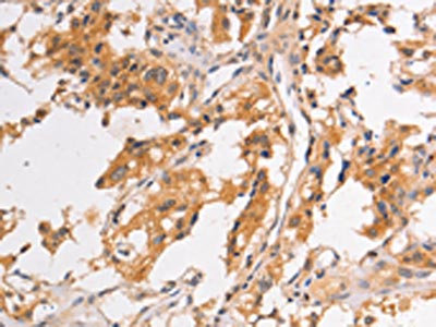
![PIP5K1A antibody [HL2011] detects PIP5K1A protein at cytoplasm and nucleus by immunohistochemical analysis. Sample: Paraffin-embedded cat spleen. PIP5K1A stained by PIP5K1A antibody [HL2011] (GTX637912) diluted at 1:100. Antigen Retrieval: Citrate buffer, pH 6.0, 15 min](https://www.genetex.com/upload/website/prouct_img/normal/GTX637912/GTX637912_44935_20230714_IHC-P_Cat_23081619_505.webp "PIP5K1A antibody [HL2011] detects PIP5K1A protein at cytoplasm and nucleus by immunohistochemical analysis. Sample: Paraffin-embedded cat spleen. PIP5K1A stained by PIP5K1A antibody [HL2011] (GTX637912) diluted at 1:100. Antigen Retrieval: Citrate buffer, pH 6.0, 15 min")
Various tissue extracts (50 μg) were separated by 7.5% SDS-PAGE, and the membrane was blotted with PIP5K1A antibody [HL2011] (GTX637912) diluted at 1:1000. The HRP-conjugated anti-rabbit IgG antibody (GTX213110-01) was used to detect the primary antibody.
PIP5K1A antibody [HL2011]
GTX637912
ApplicationsImmunoFluorescence, Western Blot, ImmunoCytoChemistry, ImmunoHistoChemistry, ImmunoHistoChemistry Paraffin
Product group Antibodies
ReactivityCanine, Feline, Human, Rat
TargetPIP5K1A
Overview
- SupplierGeneTex
- Product NamePIP5K1A antibody [HL2011]
- Delivery Days Customer9
- Application Supplier NoteWB: 1:500-1:3000. *Optimal dilutions/concentrations should be determined by the researcher.Not tested in other applications.
- ApplicationsImmunoFluorescence, Western Blot, ImmunoCytoChemistry, ImmunoHistoChemistry, ImmunoHistoChemistry Paraffin
- CertificationResearch Use Only
- ClonalityMonoclonal
- Clone IDHL2011
- Concentration1 mg/ml
- ConjugateUnconjugated
- Gene ID8394
- Target namePIP5K1A
- Target descriptionphosphatidylinositol-4-phosphate 5-kinase type 1 alpha
- Target synonymsphosphatidylinositol 4-phosphate 5-kinase type-1 alpha, 68 kDa type I phosphatidylinositol 4-phosphate 5-kinase alpha, PIP5K1-alpha, PIP5KIalpha, phosphatidylinositol 4-phosphate 5-kinase type I alpha, ptdIns(4)P-5-kinase 1 alpha
- HostRabbit
- IsotypeIgG
- Protein IDQ99755
- Protein NamePhosphatidylinositol 4-phosphate 5-kinase type-1 alpha
- Scientific DescriptionEnables 1-phosphatidylinositol-4-phosphate 5-kinase activity and kinase binding activity. Involved in several processes, including activation of GTPase activity; focal adhesion assembly; and ruffle assembly. Located in several cellular components, including lamellipodium; nuclear speck; and ruffle membrane. Colocalizes with mRNA cleavage and polyadenylation specificity factor complex. [provided by Alliance of Genome Resources, Apr 2022]
- ReactivityCanine, Feline, Human, Rat
- Storage Instruction-20°C or -80°C,2°C to 8°C
- UNSPSC41116161
Datasheet
Related products
Product group Antibodies
Anti-PIP5K1A AntibodyA36840
ApplicationsWestern Blot, ImmunoHistoChemistry
ReactivityHuman
- SizePrice
Product group Antibodies
Anti-PIP5K1A Antibody144-07941
ApplicationsImmunoFluorescence, Western Blot
ReactivityHuman, Mouse
TargetPIP5K1A
- SizePrice
Product group Antibodies

PIP5K1A AntibodyLS-C748426
ApplicationsImmunoFluorescence, Western Blot, ImmunoHistoChemistry
ReactivityHuman, Rat
TargetPIP5K1A
- SizePrice
Product group Antibodies
PIP5K1A AntibodyCSB-PA094535
ApplicationsWestern Blot, ELISA, ImmunoHistoChemistry
ReactivityHuman
TargetPIP5K1A
- SizePrice

Product group Antibodies
Anti-PIP5K1A AntibodyHPA029366
ApplicationsWestern Blot, ImmunoCytoChemistry, ImmunoHistoChemistry
ReactivityHuman
TargetPIP5K1A
- SizePrice
![Wild-type (WT) and PIP5K1A knockout (KO) 293T cell extracts (30 μg) were separated by 7.5% SDS-PAGE, and the membrane was blotted with PIP5K1A antibody [N1N3] (GTX111953) diluted at 1:2000. The HRP-conjugated anti-rabbit IgG antibody (GTX213110-01) was used to detect the primary antibody.](https://www.genetex.com/upload/website/prouct_img/normal/GTX111953/GTX111953_44853_20221209_WB_KO_watermark_22122018_357.webp)
Product group Antibodies
PIP5K1A antibody [N1N3]GTX111953
ApplicationsImmunoFluorescence, Western Blot, ImmunoCytoChemistry, ImmunoHistoChemistry, ImmunoHistoChemistry Paraffin
ReactivityHuman, Mouse
TargetPIP5K1A
- SizePrice
Product group Antibodies
PIP5K1A antibodyGTX33411
ApplicationsImmunoFluorescence, Western Blot, ImmunoCytoChemistry
ReactivityHuman
TargetPIP5K1A
- SizePrice
Product group Antibodies
Anti-PIP5K1A AntibodyCAB7941
ApplicationsWestern Blot, ELISA, ImmunoHistoChemistry, ImmunoHistoChemistry Paraffin
ReactivityHuman
TargetPIP5K1A
- SizePrice