

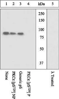
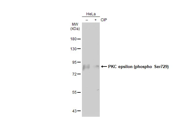
WB analysis of K562 cells stimulated with PMA using GTX25811 PKC epsilon (phospho Ser729) antibody. The membrane treated with phosphatase (Lane 5) eliminates the signal verifying that the antibody is phospho-specific.
WB analysis of K562 cells stimulated with PMA using GTX25811 PKC epsilon (phospho Ser729) antibody. The membrane treated with phosphatase (Lane 5) eliminates the signal verifying that the antibody is phospho-specific.
PKC epsilon (phospho Ser729) antibody
GTX25811
ApplicationsWestern Blot
Product group Antibodies
ReactivityHuman, Mouse
TargetPRKCE
Overview
- SupplierGeneTex
- Product NamePKC epsilon (phospho Ser729) antibody
- Delivery Days Customer9
- ApplicationsWestern Blot
- CertificationResearch Use Only
- ClonalityPolyclonal
- ConjugateUnconjugated
- Gene ID5581
- Target namePRKCE
- Target descriptionprotein kinase C epsilon
- Target synonymsPKCE, nPKC-epsilon, protein kinase C epsilon type
- HostRabbit
- IsotypeIgG
- Protein IDQ02156
- Protein NameProtein kinase C epsilon type
- Scientific DescriptionProtein kinase C (PKC) is a family of serine- and threonine-specific protein kinases that can be activated by calcium and the second messenger diacylglycerol. PKC family members phosphorylate a wide variety of protein targets and are known to be involved in diverse cellular signaling pathways. PKC family members also serve as major receptors for phorbol esters, a class of tumor promoters. Each member of the PKC family has a specific expression profile and is believed to play a distinct role in cells. The protein encoded by this gene is one of the PKC family members. This kinase has been shown to be involved in many different cellular functions, such as neuron channel activation, apoptosis, cardioprotection from ischemia, heat shock response, as well as insulin exocytosis. Knockout studies in mice suggest that this kinase is important for lipopolysaccharide (LPS)-mediated signaling in activated macrophages and may also play a role in controlling anxiety-like behavior. [provided by RefSeq, Jul 2008]
- ReactivityHuman, Mouse
- Storage Instruction-20°C or -80°C,2°C to 8°C
- UNSPSC41116161
Datasheet
Related products
Product group Antibodies
ApplicationsWestern Blot, ELISA
ReactivityHuman, Mouse, Rat
- SizePrice

Product group Antibodies
Anti-PRKCE Antibody Picoband(r)A01151-1-CARRIER-FREE
ApplicationsImmunoFluorescence, Western Blot, ELISA, ImmunoCytoChemistry
ReactivityHuman, Mouse, Rat
TargetPRKCE
- SizePrice
Product group Antibodies
Anti-PRKCE Antibody144-02110
ApplicationsWestern Blot, ImmunoCytoChemistry, ImmunoHistoChemistry
ReactivityHuman, Mouse, Rat
TargetPRKCE
- SizePrice
Product group Antibodies
References
PKC epsilon Polyclonal AntibodyBS-2329R
ApplicationsImmunoFluorescence, Western Blot, ELISA, ImmunoCytoChemistry, ImmunoHistoChemistry, ImmunoHistoChemistry Frozen, ImmunoHistoChemistry Paraffin
ReactivityBovine, Canine, Chicken, Equine, Human, Mouse, Porcine, Rabbit, Rat
TargetPRKCE
- SizePrice
Product group Antibodies
PRKCE AntibodyCSB-PA018704ESR2HU
ApplicationsImmunoPrecipitation, ELISA
ReactivityHuman
TargetPRKCE
- SizePrice
Product group Antibodies
ApplicationsImmunoPrecipitation, Western Blot, ImmunoCytoChemistry, ImmunoHistoChemistry
ReactivityPorcine, Rat
TargetPRKCE
- SizePrice
Product group Antibodies

PRKCE / PKC-Epsilon AntibodyLS-C401340
ApplicationsWestern Blot, ELISA, ImmunoHistoChemistry
ReactivityHuman, Mouse, Rat
TargetPRKCE
- SizePrice
Product group Antibodies
ApplicationsWestern Blot, ImmunoHistoChemistry, ImmunoHistoChemistry Paraffin
ReactivityHuman, Mouse, Rat
TargetPRKCE
- SizePrice

Product group Antibodies
Anti-PRKCE AntibodyHPA008236
ApplicationsImmunoCytoChemistry
ReactivityHuman
TargetPRKCE
- SizePrice
Product group Antibodies
ApplicationsWestern Blot
ReactivityHuman
TargetPRKCE
- SizePrice