

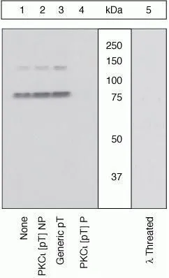
WB (peptide competition) analysis of Jurkat cells stimulated with PMA using GTX25813 PKC iota / lambda (phospho Thr563) antibody prior incubated with the non-phosphopeptide corresponding to the immunogen (Lane 2), a generic phosphothreonine-containing peptide (Lane 3), or the phosphopeptide immunogen (Lane 4). The data show that only the immunogen phosphopeptide blocks the signal, demonstrating the specificity of the antibody. The membrane treated with phosphatase (Lane 5) eliminates the signal further verifying that the antibody is phospho-specific.
WB (peptide competition) analysis of Jurkat cells stimulated with PMA using GTX25813 PKC iota / lambda (phospho Thr563) antibody prior incubated with the non-phosphopeptide corresponding to the immunogen (Lane 2), a generic phosphothreonine-containing peptide (Lane 3), or the phosphopeptide immunogen (Lane 4). The data show that only the immunogen phosphopeptide blocks the signal, demonstrating the specificity of the antibody. The membrane treated with phosphatase (Lane 5) eliminates the signal further verifying that the antibody is phospho-specific.
PKC iota / lambda (phospho Thr563) antibody
GTX25813
ApplicationsFlow Cytometry, ImmunoFluorescence, Western Blot, ImmunoCytoChemistry
Product group Antibodies
ReactivityCanine, Human, Mouse
TargetPRKCI
Overview
- SupplierGeneTex
- Product NamePKC iota / lambda (phospho Thr563) antibody
- Delivery Days Customer9
- Application Supplier NoteFACS: 1:20. *Optimal dilutions/concentrations should be determined by the researcher.Not tested in other applications.
- ApplicationsFlow Cytometry, ImmunoFluorescence, Western Blot, ImmunoCytoChemistry
- CertificationResearch Use Only
- ClonalityPolyclonal
- ConjugateUnconjugated
- Gene ID5584
- Target namePRKCI
- Target descriptionprotein kinase C iota
- Target synonymsDXS1179E, PKCI, nPKC-iota, protein kinase C iota type, PRKC-lambda/iota, aPKC-lambda/iota, atypical protein kinase C-lambda/iota
- HostRabbit
- IsotypeIgG
- Protein IDP41743
- Protein NameProtein kinase C iota type
- Scientific DescriptionThis gene encodes a member of the protein kinase C (PKC) family of serine/threonine protein kinases. The PKC family comprises at least eight members, which are differentially expressed and are involved in a wide variety of cellular processes. This protein kinase is calcium-independent and phospholipid-dependent. It is not activated by phorbolesters or diacylglycerol. This kinase can be recruited to vesicle tubular clusters (VTCs) by direct interaction with the small GTPase RAB2, where this kinase phosphorylates glyceraldehyde-3-phosphate dehydrogenase (GAPD/GAPDH) and plays a role in microtubule dynamics in the early secretory pathway. This kinase is found to be necessary for BCL-ABL-mediated resistance to drug-induced apoptosis and therefore protects leukemia cells against drug-induced apoptosis. There is a single exon pseudogene mapped on chromosome X. [provided by RefSeq, Jul 2008]
- ReactivityCanine, Human, Mouse
- Storage Instruction-20°C or -80°C,2°C to 8°C
- UNSPSC12352203
References
- Ahsan MK, Dos Reis DC, Barbieri A, et al. Loss of Serum Glucocorticoid-Inducible Kinase 1 SGK1 Worsens Malabsorption and Diarrhea in Microvillus Inclusion Disease (MVID). J Clin Med. 2022,11(14). doi: 10.3390/jcm11144179Read this paper
- Forteza R, Figueroa Y, Mashukova A, et al. Conditional knockout of polarity complex (atypical) PKCι reveals an anti-inflammatory function mediated by NF-κB. Mol Biol Cell. 2016,27(14):2186-97. doi: 10.1091/mbc.E16-02-0086Read this paper
- Kravtsov D, Mashukova A, Forteza R, et al. Myosin 5b loss of function leads to defects in polarized signaling: implication for microvillus inclusion disease pathogenesis and treatment. Am J Physiol Gastrointest Liver Physiol. 2014,307(10):G992-G1001. doi: 10.1152/ajpgi.00180.2014Read this paper
- Mashukova A, Kozhekbaeva Z, Forteza R, et al. The BAG-1 isoform BAG-1M regulates keratin-associated Hsp70 chaperoning of aPKC in intestinal cells during activation of inflammatory signaling. J Cell Sci. 2014,127(Pt 16):3568-77. doi: 10.1242/jcs.151084Read this paper
- Forteza R, Wald FA, Mashukova A, et al. Par-complex aPKC and Par3 cross-talk with innate immunity NF-κB pathway in epithelial cells. Biol Open. 2013,2(11):1264-9. doi: 10.1242/bio.20135918Read this paper
Datasheet
Related products
Product group Antibodies
ApplicationsImmunoPrecipitation, Western Blot, ImmunoCytoChemistry, ImmunoHistoChemistry
ReactivityMouse, Rat
TargetPRKCI
- SizePrice
Product group Antibodies
PKC iota Polyclonal AntibodyBS-12689R
ApplicationsImmunoFluorescence, Western Blot, ELISA, ImmunoCytoChemistry, ImmunoHistoChemistry, ImmunoHistoChemistry Frozen, ImmunoHistoChemistry Paraffin
ReactivityBovine, Equine, Human, Mouse, Porcine, Rat, Sheep
TargetPRKCI
- SizePrice
Product group Antibodies
Anti-PRKCI Antibody101-11327
ApplicationsWestern Blot, ELISA
TargetPRKCI
- SizePrice
Product group Antibodies
PRKCI AntibodyCSB-PA018707GA01HU
ApplicationsWestern Blot, ELISA, ImmunoHistoChemistry
ReactivityHuman, Mouse, Rat
TargetPRKCI
- SizePrice
Product group Antibodies

PRKCI / PKC Iota Antibody (aa375-596)LS-C373960
ApplicationsWestern Blot
ReactivityHuman
TargetPRKCI
- SizePrice

Product group Antibodies
Anti-PRKCI AntibodyHPA026574
ApplicationsWestern Blot, ImmunoCytoChemistry, ImmunoHistoChemistry
ReactivityHuman
TargetPRKCI
- SizePrice

Product group Antibodies
Anti-PKC iota/PRKCI Antibody Picoband(r)PB9321-CARRIER-FREE
ApplicationsWestern Blot, ImmunoHistoChemistry
ReactivityHuman, Mouse, Rat
TargetPRKCI
- SizePrice