

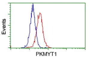
FACS analysis of HeLa cells using GTX83876 PKMYT1 antibody [1C3]. Red : Primary antibody Blue : Negative control antibody
![ICC/IF analysis of COS7 cells transiently transfected with PKMYT1 plasmid using GTX83876 PKMYT1 antibody [1C3].](https://www.genetex.com/upload/website/prouct_img/normal/GTX83876/GTX83876_857_ICCIF_w_23061420_279.webp "ICC/IF analysis of COS7 cells transiently transfected with PKMYT1 plasmid using GTX83876 PKMYT1 antibody [1C3].")
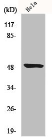
![WB analysis of MDCK and PC12 cell lysate using GTX83876 PKMYT1 antibody [1C3]. Loading : 10 ug per lane Dilution : 1:200](https://www.genetex.com/upload/website/prouct_img/normal/GTX83876/GTX83876_3466_WB_w_23061420_129.webp "WB analysis of MDCK and PC12 cell lysate using GTX83876 PKMYT1 antibody [1C3]. Loading : 10 ug per lane Dilution : 1:200")
![WB analysis of HEK293T cells transfected with PKMYT1 plasmid (Right) or empty vector (Left) for 48 hrs using GTX83876 PKMYT1 antibody [1C3]. Loading : 5 ug per lane](https://www.genetex.com/upload/website/prouct_img/normal/GTX83876/GTX83876_3966_WB_w_23061420_372.webp "WB analysis of HEK293T cells transfected with PKMYT1 plasmid (Right) or empty vector (Left) for 48 hrs using GTX83876 PKMYT1 antibody [1C3]. Loading : 5 ug per lane")
FACS analysis of HeLa cells using GTX83876 PKMYT1 antibody [1C3]. Red : Primary antibody Blue : Negative control antibody
PKMYT1 antibody [1C3]
GTX83876
ApplicationsFlow Cytometry, ImmunoFluorescence, Western Blot, ImmunoCytoChemistry
Product group Antibodies
ReactivityCanine, Human, Rat
TargetPKMYT1
Overview
- SupplierGeneTex
- Product NamePKMYT1 antibody [1C3]
- Delivery Days Customer9
- Application Supplier NoteWB: 1:1000. ICC/IF: 1:100. FCM: 1:100. *Optimal dilutions/concentrations should be determined by the researcher.Not tested in other applications.
- ApplicationsFlow Cytometry, ImmunoFluorescence, Western Blot, ImmunoCytoChemistry
- CertificationResearch Use Only
- ClonalityMonoclonal
- Clone ID1C3
- Concentration1 mg/ml
- ConjugateUnconjugated
- Gene ID9088
- Target namePKMYT1
- Target descriptionprotein kinase, membrane associated tyrosine/threonine 1
- Target synonymsMYT1, PPP1R126, membrane-associated tyrosine- and threonine-specific cdc2-inhibitory kinase, myt1 kinase, protein phosphatase 1, regulatory subunit 126
- HostMouse
- IsotypeIgG2a
- Protein IDQ99640
- Protein NameMembrane-associated tyrosine- and threonine-specific cdc2-inhibitory kinase
- Scientific DescriptionActs as a negative regulator of entry into mitosis (G2 to M transition) by phosphorylation of the CDK1 kinase specifically when CDK1 is complexed to cyclins. Mediates phosphorylation of CDK1 predominantly on Thr-14. Also involved in Golgi fragmentation. May be involved in phosphorylation of CDK1 on Tyr-15 to a lesser degree, however tyrosine kinase activity is unclear and may be indirect. May be a downstream target of Notch signaling pathway during eye development.
- ReactivityCanine, Human, Rat
- Storage Instruction-20°C or -80°C,2°C to 8°C
- UNSPSC41116161
Datasheet
Related products
Product group Antibodies
PKMYT1 AntibodyCSB-PA003356
ApplicationsWestern Blot, ELISA, ImmunoHistoChemistry
ReactivityHuman
TargetPKMYT1
- SizePrice
Product group Antibodies

MYT1 (Phospho-Ser83) AntibodyABX012617
ApplicationsELISA, ImmunoHistoChemistry
- SizePrice
Product group Antibodies
ApplicationsImmunoFluorescence, ELISA, ImmunoHistoChemistry
ReactivityHuman, Mouse, Rat
TargetPKMYT1
- SizePrice
Product group Antibodies
Anti-PKMYT1 AntibodyA306221
ApplicationsWestern Blot
ReactivityHuman, Mouse, Rat
- SizePrice

Product group Antibodies
Anti-PKMYT1 AntibodyHPA068860
ApplicationsImmunoCytoChemistry
ReactivityHuman
TargetPKMYT1
- SizePrice
Product group Antibodies

PKMYT1 Antibody (Internal)LS-C368467
ApplicationsWestern Blot, ImmunoHistoChemistry, ImmunoHistoChemistry Paraffin
ReactivityHuman, Mouse
TargetPKMYT1
- SizePrice
Product group Antibodies
PKMYT1 Polyclonal AntibodyBS-4148R
ApplicationsImmunoFluorescence, Western Blot, ELISA, ImmunoCytoChemistry, ImmunoHistoChemistry, ImmunoHistoChemistry Frozen, ImmunoHistoChemistry Paraffin
ReactivityCanine, Equine, Human, Mouse, Rat
TargetPKMYT1
- SizePrice
![WB analysis of HEK293T cells transfected with PKMYT1 plasmid (Right) or empty vector (Left) for 48 hrs using GTX83875 PKMYT1 antibody [2B4]. Loading : 5 ug per lane](https://www.genetex.com/upload/website/prouct_img/normal/GTX83875/GTX83875_3965_WB_w_23061420_688.webp)
Product group Antibodies
PKMYT1 antibody [2B4]GTX83875
ApplicationsFlow Cytometry, ImmunoFluorescence, Western Blot, ImmunoCytoChemistry
ReactivityCanine, Human, Monkey, Mouse, Rat
TargetPKMYT1
- SizePrice
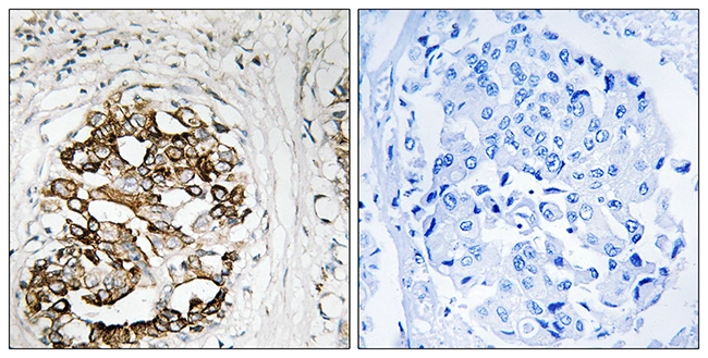
![IHC-P analysis of human endometrium tissue using GTX83877 PKMYT1 antibody [5E1]. Antigen retrieval : Heat-induced epitope retrieval by 10mM citrate buffer, pH6.0, 100oC for 10min.](https://www.genetex.com/upload/website/prouct_img/normal/GTX83877/GTX83877_1965_IHC-P_w_23061420_755.webp)
Product group Antibodies
PKMYT1 antibody [5E1]GTX83877
ApplicationsFlow Cytometry, ImmunoFluorescence, Western Blot, ImmunoCytoChemistry, ImmunoHistoChemistry, ImmunoHistoChemistry Paraffin
ReactivityHuman
TargetPKMYT1
- SizePrice
Product group Antibodies
PKMYT1 antibodyGTX87414
ApplicationsWestern Blot, ImmunoHistoChemistry, ImmunoHistoChemistry Paraffin
ReactivityHuman
TargetPKMYT1
- SizePrice