

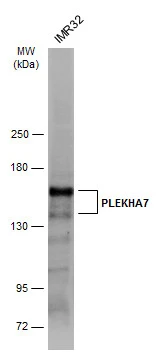
Whole cell extract (30 μg) was separated by 5% SDS-PAGE, and the membrane was blotted with PLEKHA7 antibody (GTX131146) diluted at 1:500. The HRP-conjugated anti-rabbit IgG antibody (GTX213110-01) was used to detect the primary antibody.
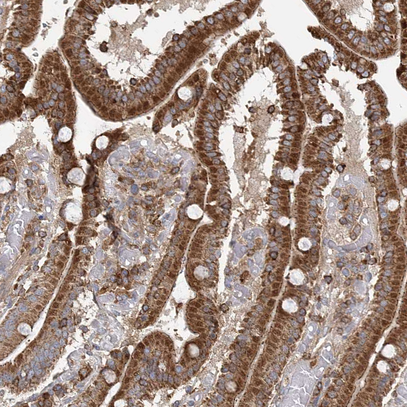
![PLEKHA7 antibody detects PLEKHA7 protein expression by immunohistochemical analysis. Sample: Paraffin-Embedded mouse colon. Green: PLEKHA7 stained by PLEKHA7 antibody (GTX131146) diluted at 1:1000. Red: E-Cadherin, stained byE-Cadherin antibody [GT477] (GTX629691) diluted at 1:250. Blue: Fluoroshield with DAPI (GTX30920).
Antigen Retrieval: Citrate buffer, pH 6.0, 15 min](https://www.genetex.com/upload/website/prouct_img/normal/GTX131146/GTX131146_42718_20170428_IHC-P_M_w_23060523_850.webp "PLEKHA7 antibody detects PLEKHA7 protein expression by immunohistochemical analysis. Sample: Paraffin-Embedded mouse colon. Green: PLEKHA7 stained by PLEKHA7 antibody (GTX131146) diluted at 1:1000. Red: E-Cadherin, stained byE-Cadherin antibody [GT477] (GTX629691) diluted at 1:250. Blue: Fluoroshield with DAPI (GTX30920).
Antigen Retrieval: Citrate buffer, pH 6.0, 15 min")
 were separated by 5% SDS-PAGE, and the membrane was blotted with PLEKHA7 antibody (GTX131146) diluted at 1:500. The HRP-conjugated anti-rabbit IgG antibody (GTX213110-01) was used to detect the primary antibody.")
Whole cell extract (30 μg) was separated by 5% SDS-PAGE, and the membrane was blotted with PLEKHA7 antibody (GTX131146) diluted at 1:500. The HRP-conjugated anti-rabbit IgG antibody (GTX213110-01) was used to detect the primary antibody.
PLEKHA7 antibody
GTX131146
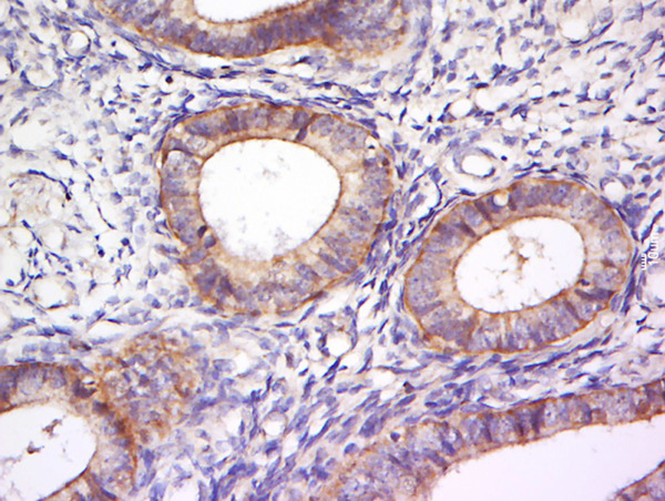
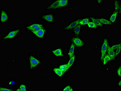
ApplicationsWestern Blot, ImmunoHistoChemistry, ImmunoHistoChemistry Paraffin
Product group Antibodies
ReactivityHuman, Mouse
TargetPLEKHA7
Overview
- SupplierGeneTex
- Product NamePLEKHA7 antibody
- Delivery Days Customer9
- Application Supplier NoteWB: 1:500-1:3000. IHC-P: 1:100-1:1000. *Optimal dilutions/concentrations should be determined by the researcher.Not tested in other applications.
- ApplicationsWestern Blot, ImmunoHistoChemistry, ImmunoHistoChemistry Paraffin
- CertificationResearch Use Only
- ClonalityPolyclonal
- Concentration0.91 mg/ml
- ConjugateUnconjugated
- Gene ID144100
- Target namePLEKHA7
- Target descriptionpleckstrin homology domain containing A7
- Target synonymspleckstrin homology domain-containing family A member 7, PH domain-containing family A member 7, pleckstrin homology domain containing, family A member 2, pleckstrin homology domain containing, family A member 7, pleckstrin homology domain-containing family A member 6
- HostRabbit
- IsotypeIgG
- Protein IDQ6IQ23
- Protein NamePleckstrin homology domain-containing family A member 7
- Scientific DescriptionRequired for zonula adherens biogenesis and maintenance. Acts via its interaction with KIAA1543/Nezha, which anchors microtubules at their minus-ends to zonula adherens, leading to recruit KIFC3 kinesin to junctional site.
- ReactivityHuman, Mouse
- Storage Instruction-20°C or -80°C,2°C to 8°C
- UNSPSC41116161
Datasheet
Related products

Product group Antibodies
PLEKHA7 Polyclonal AntibodyBS-13730R
ApplicationsImmunoFluorescence, Western Blot, ELISA, ImmunoCytoChemistry, ImmunoHistoChemistry, ImmunoHistoChemistry Frozen, ImmunoHistoChemistry Paraffin
ReactivityHuman
TargetPLEKHA7
- SizePrice
Product group Antibodies
PLEKHA7 AntibodyCSB-PA750339LA01HU
ApplicationsImmunoFluorescence, ELISA
ReactivityHuman
TargetPLEKHA7
- SizePrice
Product group Antibodies

PLEKHA7 Antibody (HRP)LS-C501106
ApplicationsELISA
ReactivityHuman
TargetPLEKHA7
- SizePrice
Product group Antibodies
Anti-PLEKHA7 AntibodyHPA038610
ApplicationsWestern Blot, ImmunoCytoChemistry, ImmunoHistoChemistry
ReactivityHuman
TargetPLEKHA7
- SizePrice