

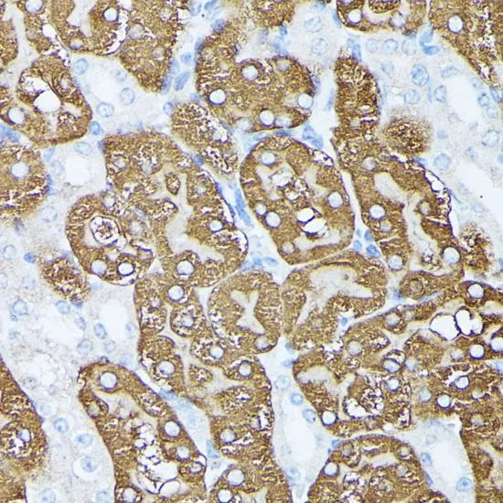
IHC-P analysis of mouse kidney tissue using GTX66355 PLS1 antibody. Dilution : 1:100


IHC-P analysis of mouse kidney tissue using GTX66355 PLS1 antibody. Dilution : 1:100
PLS1 antibody
GTX66355
ApplicationsWestern Blot, ImmunoHistoChemistry, ImmunoHistoChemistry Paraffin
Product group Antibodies
ReactivityHuman, Mouse, Rat
TargetPLS1
Overview
- SupplierGeneTex
- Product NamePLS1 antibody
- Delivery Days Customer9
- Application Supplier NoteWB: 1:200 - 1:2000. IHC-P: 1:50 - 1:200. *Optimal dilutions/concentrations should be determined by the researcher.Not tested in other applications.
- ApplicationsWestern Blot, ImmunoHistoChemistry, ImmunoHistoChemistry Paraffin
- CertificationResearch Use Only
- ClonalityPolyclonal
- ConjugateUnconjugated
- Gene ID5357
- Target namePLS1
- Target descriptionplastin 1
- Target synonymsDFNA76, plastin-1, I plastin, fimbrin, intestine specific plastin
- HostRabbit
- IsotypeIgG
- Protein IDQ14651
- Protein NamePlastin-1
- Scientific DescriptionPlastins are a family of actin-binding proteins that are conserved throughout eukaryote evolution and expressed in most tissues of higher eukaryotes. In humans, two ubiquitous plastin isoforms (L and T) have been identified. The protein encoded by this gene is a third distinct plastin isoform, which is specifically expressed at high levels in the small intestine. Alternatively spliced transcript variants varying in the 5 UTR, but encoding the same protein, have been found for this gene. A pseudogene of this gene is found on chromosome 11.[provided by RefSeq, Feb 2010]
- ReactivityHuman, Mouse, Rat
- Storage Instruction-20°C or -80°C,2°C to 8°C
- UNSPSC41116161
Datasheet
Related products
Product group Antibodies
Anti-PLS1 AntibodyA90953
ApplicationsWestern Blot, ImmunoHistoChemistry
ReactivityHuman, Mouse, Rat
- SizePrice

Product group Antibodies
Anti-PLS1 Antibody Picoband(r)A07675-1-CARRIER-FREE
ApplicationsFlow Cytometry, ImmunoFluorescence, Western Blot, ELISA, ImmunoCytoChemistry, ImmunoHistoChemistry
ReactivityHuman, Mouse, Rat
TargetPLS1
- SizePrice
Product group Antibodies
Anti-PLS1 Antibody144-60987
ApplicationsWestern Blot
ReactivityHuman, Mouse, Rat
TargetPLS1
- SizePrice
Product group Antibodies

PLS1 / Fimbrin AntibodyLS-C750274
ApplicationsWestern Blot
ReactivityHuman, Mouse, Rat
TargetPLS1
- SizePrice
Product group Antibodies
PLS1 AntibodyCSB-PA623928LA01HU
ApplicationsImmunoFluorescence, ImmunoPrecipitation, Western Blot, ELISA, ImmunoHistoChemistry
ReactivityHuman
TargetPLS1
- SizePrice
Product group Antibodies
Pls1 Polyclonal AntibodyCAC11500
ApplicationsImmunoFluorescence, ImmunoPrecipitation, Western Blot, ELISA, ImmunoHistoChemistry
TargetPLS1
- SizePrice

Product group Antibodies
Anti-PLS1 AntibodyHPA055744
ApplicationsImmunoHistoChemistry
ReactivityHuman
TargetPLS1
- SizePrice
Product group Antibodies
PLS1 antibody, N-termGTX45058
ApplicationsWestern Blot
ReactivityHuman
TargetPLS1
- SizePrice
Product group Antibodies
Anti-PLS1 AntibodyCAB15303
ApplicationsWestern Blot, ELISA, ImmunoHistoChemistry, ImmunoHistoChemistry Paraffin
ReactivityHuman
TargetPLS1
- SizePrice