

![Pmel17 / gp100 / SILV(HMB45), CF405S conjugate, 0.1mg/mL [26628-22-8]](https://biotium.com/wp-content/uploads/2016/12/BNUB0444-1-1.jpg "Pmel17 / gp100 / SILV(HMB45), CF405S conjugate, 0.1mg/mL [26628-22-8]")
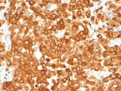
Pmel17 / gp100 / SILV(HMB45), CF405S conjugate, 0.1mg/mL [26628-22-8]
BNC040444
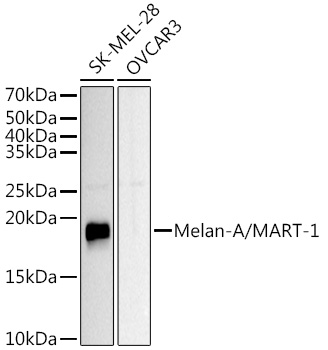
ApplicationsWestern Blot, ImmunoHistoChemistry, ImmunoHistoChemistry Paraffin
Product group Antibodies
TargetPMEL
Overview
- SupplierBiotium
- Product NamePmel17 / gp100 / SILV(HMB45), CF405S conjugate, 0.1mg/mL [26628-22-8]
- Delivery Days Customer9
- ApplicationsWestern Blot, ImmunoHistoChemistry, ImmunoHistoChemistry Paraffin
- CertificationResearch Use Only
- ClonalityMonoclonal
- Clone IDHMB45
- Concentration0.1 mg/ml
- ConjugateOther Conjugate
- Gene ID6490
- Target namePMEL
- Target descriptionpremelanosome protein
- Target synonymsD12S53E, HMB-45, HMB45, ME20, ME20-M, ME20M, P1, P100, PMEL17, SI, SIL, SILV, gp100, melanocyte protein PMEL, melanocyte protein Pmel 17, melanocyte protein mel 17, melanocytes lineage-specific antigen GP100, melanoma-associated ME20 antigen, melanosomal matrix protein17, silver locus protein homolog, silver, mouse, homolog of
- HostMouse
- IsotypeIgG1
- Protein IDP40967
- Protein NameMelanocyte protein PMEL
- Scientific DescriptionBy immunohistochemistry, this antibody specifically recognizes a protein in melanocytes and melanomas. It reacts with junctional and blue nevus cells and variably with fetal and neonatal melanocytes. Intradermal nevi, normal adult melanocytes, and non-melanocytic cells are negative. It does not stain tumor cells of epithelial, lymphoid, glial, or mesenchymal origin. Metastatic amelanotic melanoma can often be confused with a variety of poorly differentiated carcinomas, large cell lymphomas, and sarcomas using H & E stains alone. It is also difficult to differentiate melanoma from spindle cell carcinomas and various types of mesenchymal neoplasms. This MAb stains fetal and neonatal melanocytes, junctional and blue nevus cells, malignant melanoma, and angiomyolipoma (PEComa).Primary antibodies are available purified, or with a selection of fluorescent CF® Dyes and other labels. CF® Dyes offer exceptional brightness and photostability. Note: Conjugates of blue fluorescent dyes like CF®405S and CF®405M are not recommended for detecting low abundance targets, because blue dyes have lower fluorescence and can give higher non-specific background than other dye colors.
- SourceAnimal
- Storage Instruction2°C to 8°C
- UNSPSC12352203
Related products
Product group Antibodies
PMEL Recombinant AntibodyBSM-61122R
ApplicationsImmunoFluorescence, Western Blot, ImmunoHistoChemistry, ImmunoHistoChemistry Frozen, ImmunoHistoChemistry Paraffin
TargetPMEL
- SizePrice
Product group Antibodies
Pmel Polyclonal AntibodyCAC08845
ApplicationsWestern Blot, ELISA, ImmunoHistoChemistry
TargetPMEL
- SizePrice
Product group Antibodies
Anti-Melanoma AntibodyA15151
ApplicationsImmunoFluorescence, Western Blot, ImmunoCytoChemistry
- SizePrice

Product group Antibodies
References

ApplicationsImmunoFluorescence, Western Blot, ELISA
TargetPMEL
- SizePrice
Product group Antibodies
ApplicationsImmunoHistoChemistry, ImmunoHistoChemistry Paraffin
TargetPMEL
- SizePrice
Product group Antibodies

Melanoma, HMB45ME505
ApplicationsImmunoHistoChemistry, ImmunoHistoChemistry Paraffin
TargetPMEL
- SizePrice