

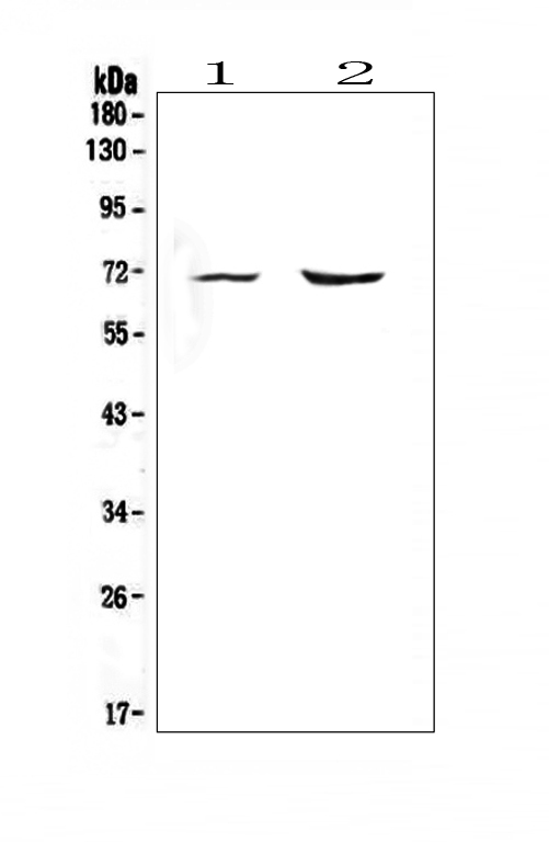
Figure 1. Western blot analysis of Melanoma gp100 using anti-Melanoma gp100 antibody (A01262-1). Electrophoresis was performed on a 5-20% SDS-PAGE gel at 70V (Stacking gel) / 90V (Resolving gel) for 2-3 hours. The sample well of each lane was loaded with 50ug of sample under reducing conditions. Lane 1: rat heart tissue lysate, Lane 2: mouse heart tissue lysate. After Electrophoresis, proteins were transferred to a Nitrocellulose membrane at 150mA for 50-90 minutes. Blocked the membrane with 5% Non-fat Milk/ TBS for 1.5 hour at RT. The membrane was incubated with rabbit anti-Melanoma gp100 antigen affinity purified polyclonal antibody (Catalog # A01262-1) at 0.5 microg/mL overnight at 4°C, then washed with TBS-0.1%Tween 3 times with 5 minutes each and probed with a goat anti-rabbit IgG-HRP secondary antibody at a dilution of 1:10000 for 1.5 hour at RT. The signal is developed using an Enhanced Chemiluminescent detection (ECL) kit (Catalog # EK1002) with Tanon 5200 system. A specific band was detected for Melanoma gp100 at approximately 70KD. The expected band size for Melanoma gp100 is at 70KD.
Figure 1. Western blot analysis of Melanoma gp100 using anti-Melanoma gp100 antibody (A01262-1). Electrophoresis was performed on a 5-20% SDS-PAGE gel at 70V (Stacking gel) / 90V (Resolving gel) for 2-3 hours. The sample well of each lane was loaded with 50ug of sample under reducing conditions. Lane 1: rat heart tissue lysate, Lane 2: mouse heart tissue lysate. After Electrophoresis, proteins were transferred to a Nitrocellulose membrane at 150mA for 50-90 minutes. Blocked the membrane with 5% Non-fat Milk/ TBS for 1.5 hour at RT. The membrane was incubated with rabbit anti-Melanoma gp100 antigen affinity purified polyclonal antibody (Catalog # A01262-1) at 0.5 microg/mL overnight at 4°C, then washed with TBS-0.1%Tween 3 times with 5 minutes each and probed with a goat anti-rabbit IgG-HRP secondary antibody at a dilution of 1:10000 for 1.5 hour at RT. The signal is developed using an Enhanced Chemiluminescent detection (ECL) kit (Catalog # EK1002) with Tanon 5200 system. A specific band was detected for Melanoma gp100 at approximately 70KD. The expected band size for Melanoma gp100 is at 70KD.
Anti-Melanoma gp100/PMEL Antibody Picoband(r)
A01262-1-CARRIER-FREE
ApplicationsWestern Blot
Product group Antibodies
ReactivityHuman, Mouse, Rat
TargetPMEL
Overview
- SupplierBoster Bio
- Product NameAnti-Melanoma gp100/PMEL Antibody Picoband(r)
- Delivery Days Customer9
- ApplicationsWestern Blot
- CertificationResearch Use Only
- ClonalityPolyclonal
- Concentration500 ug/ml
- Gene ID6490
- Target namePMEL
- Target descriptionpremelanosome protein
- Target synonymsD12S53E, HMB-45, HMB45, ME20, ME20-M, ME20M, P1, P100, PMEL17, SI, SIL, SILV, gp100, melanocyte protein PMEL, melanocyte protein Pmel 17, melanocyte protein mel 17, melanocytes lineage-specific antigen GP100, melanoma-associated ME20 antigen, melanosomal matrix protein17, silver locus protein homolog, silver, mouse, homolog of
- HostRabbit
- IsotypeIgG
- Protein IDP40967
- Protein NameMelanocyte protein PMEL
- Scientific DescriptionBoster Bio Anti-Melanoma gp100/PMEL Antibody Picoband® catalog # A01262-1. Tested in WB applications. This antibody reacts with Human, Mouse, Rat. The brand Picoband indicates this is a premium antibody that guarantees superior quality, high affinity, and strong signals with minimal background in Western blot applications. Only our best-performing antibodies are designated as Picoband, ensuring unmatched performance.
- ReactivityHuman, Mouse, Rat
- Storage Instruction-20°C,2°C to 8°C
- UNSPSC12352203
Related products
Product group Antibodies
Anti-Melanoma AntibodyA15151
ApplicationsImmunoFluorescence, Western Blot, ImmunoCytoChemistry
ReactivityHuman, Mouse, Rat
- SizePrice
Product group Antibodies
Anti-gp100 Antibody188-10170
ApplicationsWestern Blot
ReactivityCanine, Human, Rat
TargetPMEL
- SizePrice

Product group Antibodies

PMEL / SILV / gp100 AntibodyLS-C761105
ApplicationsWestern Blot
ReactivityHuman, Mouse, Rat
TargetPMEL
- SizePrice
Product group Antibodies
PMEL Recombinant AntibodyBSM-61122R
ApplicationsImmunoFluorescence, Western Blot, ImmunoHistoChemistry, ImmunoHistoChemistry Frozen, ImmunoHistoChemistry Paraffin
TargetPMEL
- SizePrice
Product group Antibodies
References

ApplicationsImmunoFluorescence, Western Blot, ELISA
ReactivityHuman, Mouse
TargetPMEL
- SizePrice
Product group Antibodies
Pmel Polyclonal AntibodyCAC08845
ApplicationsWestern Blot, ELISA, ImmunoHistoChemistry
TargetPMEL
- SizePrice
Product group Antibodies
PMEL AntibodyCSB-PA021324LA01HU
ApplicationsWestern Blot, ELISA, ImmunoHistoChemistry
ReactivityHuman
TargetPMEL
- SizePrice
Product group Antibodies

Melanoma, HMB45ME505
ApplicationsImmunoHistoChemistry, ImmunoHistoChemistry Paraffin
ReactivityHuman
TargetPMEL
- SizePrice