

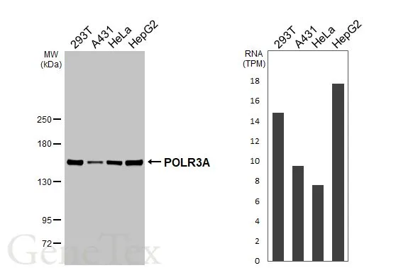
Various whole cell extracts (30 μg) were separated by 5% SDS-PAGE, and the membrane was blotted with POLR3A antibody [HL3437] (GTX641310) diluted at 1:1000. The HRP-conjugated anti-rabbit IgG antibody (GTX213110-01) was used to detect the primary antibody. Corresponding RNA expression data for the same cell lines are based on Human Protein Atlas program.
![Various whole cell extracts (30 μg) were separated by 5% SDS-PAGE, and the membrane was blotted with POLR3A antibody [HL3437] (GTX641310) diluted at 1:1000. The HRP-conjugated anti-rabbit IgG antibody (GTX213110-01) was used to detect the primary antibody.](https://www.genetex.com/upload/website/prouct_img/normal/GTX641310/GTX641310_T-45593_20241115_WB_M_R_24111918_833.webp "Various whole cell extracts (30 μg) were separated by 5% SDS-PAGE, and the membrane was blotted with POLR3A antibody [HL3437] (GTX641310) diluted at 1:1000. The HRP-conjugated anti-rabbit IgG antibody (GTX213110-01) was used to detect the primary antibody.")
![POLR3A antibody [HL3437] detects POLR3A protein by immunohistochemical analysis. Sample: Paraffin-embedded rat tissues. POLR3A stained by POLR3A antibody [HL3437] (GTX641310) diluted at 1:200. Antigen Retrieval: Citrate buffer, pH 6.0, 15 min Corresponding RNA levels (RPKM) in the tissues are based on NCBI database.](https://www.genetex.com/upload/website/prouct_img/normal/GTX641310/GTX641310_T-45593_20250113_IHC-P_R_Multiple_RPKM_25020422_157.webp "POLR3A antibody [HL3437] detects POLR3A protein by immunohistochemical analysis. Sample: Paraffin-embedded rat tissues. POLR3A stained by POLR3A antibody [HL3437] (GTX641310) diluted at 1:200. Antigen Retrieval: Citrate buffer, pH 6.0, 15 min Corresponding RNA levels (RPKM) in the tissues are based on NCBI database.")
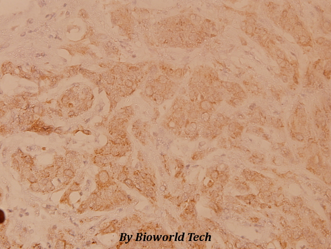
![POLR3A antibody [HL3437] detects POLR3A protein by immunohistochemical analysis. Sample: Paraffin-embedded human tissues. POLR3A stained by POLR3A antibody [HL3437] (GTX641310) diluted at 1:200. Antigen Retrieval: Citrate buffer, pH 6.0, 15 min Corresponding RNA levels (RPKM) in the tissues are based on NCBI database.](https://www.genetex.com/upload/website/prouct_img/normal/GTX641310/GTX641310_T-45593_20250113_IHC-P_Multiple_RPKM_25020422_775.webp "POLR3A antibody [HL3437] detects POLR3A protein by immunohistochemical analysis. Sample: Paraffin-embedded human tissues. POLR3A stained by POLR3A antibody [HL3437] (GTX641310) diluted at 1:200. Antigen Retrieval: Citrate buffer, pH 6.0, 15 min Corresponding RNA levels (RPKM) in the tissues are based on NCBI database.")
![POLR3A antibody [HL3437] detects POLR3A protein by immunohistochemical analysis. Sample: Paraffin-embedded mouse tissues. POLR3A stained by POLR3A antibody [HL3437] (GTX641310) diluted at 1:200. Antigen Retrieval: Citrate buffer, pH 6.0, 15 min](https://www.genetex.com/upload/website/prouct_img/normal/GTX641310/GTX641310_T-45593_20250214_IHC-P_M_Multiple_RPKM_25031220_978.webp "POLR3A antibody [HL3437] detects POLR3A protein by immunohistochemical analysis. Sample: Paraffin-embedded mouse tissues. POLR3A stained by POLR3A antibody [HL3437] (GTX641310) diluted at 1:200. Antigen Retrieval: Citrate buffer, pH 6.0, 15 min")
![POLR3A antibody [HL3437] detects POLR3A protein by immunofluorescent analysis. Sample: HepG2 cells were fixed in ice-cold MeOH for 5 min. Green: POLR3A stained by POLR3A antibody [HL3437] (GTX641310) diluted at 1:500. Red: alpha Tubulin, a cytoskeleton marker, stained by alpha Tubulin antibody [GT114] (GTX628802) diluted at 1:1000.](https://www.genetex.com/upload/website/prouct_img/normal/GTX641310/GTX641310_45663_20250425_ICC_IF_25052800_869.webp "POLR3A antibody [HL3437] detects POLR3A protein by immunofluorescent analysis. Sample: HepG2 cells were fixed in ice-cold MeOH for 5 min. Green: POLR3A stained by POLR3A antibody [HL3437] (GTX641310) diluted at 1:500. Red: alpha Tubulin, a cytoskeleton marker, stained by alpha Tubulin antibody [GT114] (GTX628802) diluted at 1:1000.")
![Whole zebrafish extract (30 μg) was separated by 5% SDS-PAGE, and the membrane was blotted with POLR3A antibody [HL3437] (GTX641310) diluted at 1:1000. The HRP-conjugated anti-rabbit IgG antibody (GTX213110-01) was used to detect the primary antibody.](https://www.genetex.com/upload/website/prouct_img/normal/GTX641310/GTX641310_45663_20250530_WB_Z_25061003_756.webp "Whole zebrafish extract (30 μg) was separated by 5% SDS-PAGE, and the membrane was blotted with POLR3A antibody [HL3437] (GTX641310) diluted at 1:1000. The HRP-conjugated anti-rabbit IgG antibody (GTX213110-01) was used to detect the primary antibody.")
![Whole Japanese medaka extract (30 μg) was separated by 5% SDS-PAGE, and the membrane was blotted with POLR3A antibody [HL3437] (GTX641310) diluted at 1:1000. The HRP-conjugated anti-rabbit IgG antibody (GTX213110-01) was used to detect the primary antibody, and the signal was developed with Trident femto Western HRP Substrate.](https://www.genetex.com/upload/website/prouct_img/normal/GTX641310/GTX641310_45663_20251003_WB_JapaneseMedaka_25100823_422.webp "Whole Japanese medaka extract (30 μg) was separated by 5% SDS-PAGE, and the membrane was blotted with POLR3A antibody [HL3437] (GTX641310) diluted at 1:1000. The HRP-conjugated anti-rabbit IgG antibody (GTX213110-01) was used to detect the primary antibody, and the signal was developed with Trident femto Western HRP Substrate.")
Various whole cell extracts (30 μg) were separated by 5% SDS-PAGE, and the membrane was blotted with POLR3A antibody [HL3437] (GTX641310) diluted at 1:1000. The HRP-conjugated anti-rabbit IgG antibody (GTX213110-01) was used to detect the primary antibody. Corresponding RNA expression data for the same cell lines are based on Human Protein Atlas program.
POLR3A antibody [HL3437]
GTX641310
ApplicationsImmunoFluorescence, Western Blot, ImmunoCytoChemistry, ImmunoHistoChemistry, ImmunoHistoChemistry Paraffin
Product group Antibodies
ReactivityHuman, Mouse, Rat
TargetPOLR3A
Overview
- SupplierGeneTex
- Product NamePOLR3A antibody [HL3437]
- Delivery Days Customer9
- Application Supplier NoteWB: 1:500-1:3000. *Optimal dilutions/concentrations should be determined by the researcher.Not tested in other applications.
- ApplicationsImmunoFluorescence, Western Blot, ImmunoCytoChemistry, ImmunoHistoChemistry, ImmunoHistoChemistry Paraffin
- CertificationResearch Use Only
- ClonalityMonoclonal
- Clone IDHL3437
- Concentration1 mg/ml
- ConjugateUnconjugated
- Gene ID11128
- Target namePOLR3A
- Target descriptionRNA polymerase III subunit A
- Target synonymsADDH, C160, HLD7, RPC1, RPC155, WDRTS, hRPC155, DNA-directed RNA polymerase III subunit RPC1, DNA-directed RNA polymerase III largest subunit, DNA-directed RNA polymerase III subunit A, RNA polymerase III 155 kDa subunit, RNA polymerase III subunit C1, RNA polymerase III subunit C160, RNA polymerase III subunit RPC155-D, polymerase (RNA) III (DNA directed) polypeptide A, 155kDa, polymerase (RNA) III subunit A
- HostRabbit
- IsotypeIgG
- Protein IDO14802
- Protein NameDNA-directed RNA polymerase III subunit RPC1
- Scientific DescriptionThe protein encoded by this gene is the catalytic component of RNA polymerase III, which synthesizes small RNAs. The encoded protein also acts as a sensor to detect foreign DNA and trigger an innate immune response. [provided by RefSeq, Aug 2011]
- ReactivityHuman, Mouse, Rat
- Storage Instruction-20°C or -80°C,2°C to 8°C
- UNSPSC41116161
Related products
Product group Antibodies
POLR3A AntibodyCSB-PA003829
ApplicationsWestern Blot, ELISA, ImmunoHistoChemistry
ReactivityHuman, Monkey, Mouse
TargetPOLR3A
- SizePrice

Product group Antibodies
ApplicationsImmunoFluorescence, Western Blot, ELISA, ImmunoCytoChemistry, ImmunoHistoChemistry
TargetPOLR3A
- SizePrice
Product group Antibodies
ApplicationsWestern Blot, ImmunoHistoChemistry
ReactivityHuman, Mouse
- SizePrice

Product group Antibodies
Anti-POLR3A AntibodyHPA037926
ApplicationsWestern Blot, ChIP Chromatin ImmunoPrecipitation, ImmunoCytoChemistry
ReactivityHuman
TargetPOLR3A
- SizePrice
Product group Antibodies

POLR3A Antibody (N-Terminus)LS-C368898
ApplicationsWestern Blot, ImmunoHistoChemistry, ImmunoHistoChemistry Paraffin
ReactivityBovine, Canine, Chicken, Human, Monkey, Mouse, Porcine, Rabbit, Rat, Zebra Fish
TargetPOLR3A
- SizePrice
Product group Antibodies
POLR3A Polyclonal AntibodyCAC13164
ApplicationsChIP Chromatin ImmunoPrecipitation, ELISA, ImmunoHistoChemistry
TargetPOLR3A
- SizePrice
Product group Antibodies
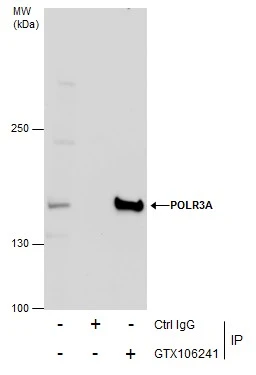
POLR3A antibodyGTX106241
ApplicationsImmunoFluorescence, ImmunoPrecipitation, Western Blot, ImmunoCytoChemistry
ReactivityHuman, Mouse, Rat
TargetPOLR3A
- SizePrice
Product group Antibodies
Anti-POLR3A Antibody107-10593
ApplicationsImmunoFluorescence, Western Blot, ImmunoCytoChemistry
ReactivityHuman
TargetPOLR3A
- SizePrice