

Primary Antibodies
Product group Antibodies
Mono-methyl-HIST1H2AG (K9) AntibodyCSB-PA010389OA09ME1HU
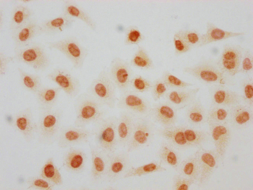
ApplicationsImmunoFluorescence, ChIP Chromatin ImmunoPrecipitation, ELISA, ImmunoCytoChemistry
ReactivityHuman
TargetH2AC13
- SizePrice
Product group Antibodies
HIST1H2AG (Ab-9) AntibodyCSB-PA010389OA09NACHU
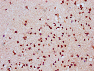
ApplicationsImmunoFluorescence, ELISA, ImmunoHistoChemistry
ReactivityHuman
TargetH2AC13
- SizePrice
Product group Antibodies
HIST1H2AG (Ab-9) AntibodyCSB-PA010389OA09NBHBHU
ApplicationsELISA, ImmunoHistoChemistry
ReactivityHuman
TargetH2AC13
- SizePrice
Product group Antibodies
HIST1H2AG (Ab-9) AntibodyCSB-PA010389OA09NHIBHU
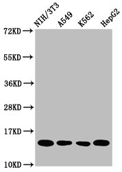
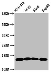
ApplicationsImmunoFluorescence, Western Blot, ELISA, ImmunoHistoChemistry
ReactivityHuman
TargetH2AC13
- SizePrice
Product group Antibodies
HIST1H2AG (Ab-9) AntibodyCSB-PA010389OA09NME1HU
ApplicationsChIP Chromatin ImmunoPrecipitation, ELISA
ReactivityHuman
TargetH2AC13
- SizePrice
Product group Antibodies
HIST1H2AG (Ab-9) AntibodyCSB-PA010389OA09NSUCHU
ApplicationsImmunoFluorescence, Western Blot, ELISA, ImmunoHistoChemistry
ReactivityHuman
TargetH2AC13
- SizePrice
Product group Antibodies
Formyl-HIST1H2AG (K118) AntibodyCSB-PA010389OA118FORHU
ApplicationsImmunoFluorescence, Western Blot, ELISA
ReactivityHuman
TargetH2AC13
- SizePrice
Product group Antibodies
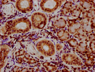
HIST1H2AG (Ab-118) AntibodyCSB-PA010389OA118NBHBHU
ApplicationsELISA, ImmunoHistoChemistry
ReactivityHuman
TargetH2AC13
- SizePrice
Product group Antibodies
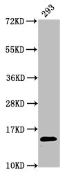
HIST1H2AG (Ab-118) AntibodyCSB-PA010389OA118NFORHU
ApplicationsImmunoFluorescence, ImmunoPrecipitation, Western Blot, ELISA, ImmunoHistoChemistry
ReactivityHuman, Mouse
TargetH2AC13
- SizePrice
Product group Antibodies
HIST1H2AG (Ab-118) AntibodyCSB-PA010389OA118NHIBHU
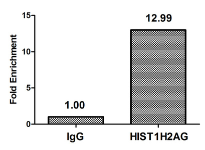
ApplicationsImmunoFluorescence, ChIP Chromatin ImmunoPrecipitation, ELISA, ImmunoHistoChemistry
ReactivityHuman
TargetH2AC13
- SizePrice
Product group Antibodies
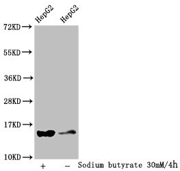
HIST1H2AG (Ab-118) AntibodyCSB-PA010389OA118NME1HU
ApplicationsImmunoPrecipitation, Western Blot, ChIP Chromatin ImmunoPrecipitation, ELISA
ReactivityHuman, Mouse
TargetH2AC13
- SizePrice
Product group Antibodies
Crotonyl-HIST1H2AG (K119) AntibodyCSB-PA010389OA119CRHU
ApplicationsELISA, ImmunoCytoChemistry
ReactivityHuman
TargetH2AC13
- SizePrice
Didn't find what you were looking for?
Search through our product groups to find the right product
Back to overview