

IHC image of CSB-PA018746LA01HU diluted at 1:200 and staining in paraffin-embedded human placenta tissue performed on a Leica BondTM system. After dewaxing and hydration, antigen retrieval was mediated by high pressure in a citrate buffer (pH 6.0). Section was blocked with 10% normal goat serum 30min at RT. Then primary antibody (1% BSA) was incubated at 4°C overnight. The primary is detected by a biotinylated secondary antibody and visualized using an HRP conjugated SP system.
. Section was blocked with 10% normal goat serum 30min at RT. Then primary antibody (1% BSA) was incubated at 4°C overnight. The primary is detected by a biotinylated secondary antibody and visualized using an HRP conjugated SP system.")
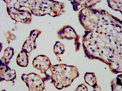
IHC image of CSB-PA018746LA01HU diluted at 1:200 and staining in paraffin-embedded human placenta tissue performed on a Leica BondTM system. After dewaxing and hydration, antigen retrieval was mediated by high pressure in a citrate buffer (pH 6.0). Section was blocked with 10% normal goat serum 30min at RT. Then primary antibody (1% BSA) was incubated at 4°C overnight. The primary is detected by a biotinylated secondary antibody and visualized using an HRP conjugated SP system.
PROK1 Antibody
CSB-PA018746LA01HU
ApplicationsELISA, ImmunoHistoChemistry
Product group Antibodies
ReactivityHuman
TargetPROK1
Overview
- SupplierCusabio
- Product NamePROK1 Antibody
- Delivery Days Customer20
- ApplicationsELISA, ImmunoHistoChemistry
- CertificationResearch Use Only
- ClonalityPolyclonal
- ConjugateUnconjugated
- Gene ID84432
- Target namePROK1
- Target descriptionprokineticin 1
- Target synonymsEGVEGF, PK1, PRK1, prokineticin-1, EG-VEGF, black mamba toxin-related protein, endocrine-gland-derived vascular endothelial growth factor, mambakine
- HostRabbit
- IsotypeIgG
- Protein IDP58294
- Protein NameProkineticin-1
- Scientific DescriptionPotently contracts gastrointestinal (GI) smooth muscle. Induces proliferation, migration and fenestration (the formation of membrane discontinuities) in capillary endothelial cells derived from endocrine glands. Has little or no effect on a variety of other endothelial and non-endothelial cell types. Induces proliferation and differentiation, but not migration, of enteric neural crest cells. Directly influences neuroblastoma progression by promoting the proliferation and migration of neuroblastoma cells. Positively regulates PTGS2 expression and prostaglandin synthesis. May play a role in placentation. May play a role in normal and pathological testis angiogenesis.
- ReactivityHuman
- Storage Instruction-20°C or -80°C
- UNSPSC41116161
Related products
Product group Antibodies
Prokineticin 1 Recombinant AntibodyBSM-62397R
ApplicationsFlow Cytometry, Western Blot
ReactivityHuman, Mouse
TargetPROK1
- SizePrice
Product group Antibodies
ApplicationsImmunoPrecipitation, Western Blot, ImmunoCytoChemistry, ImmunoHistoChemistry
TargetPROK1
- SizePrice
![IHC-P analysis of formalin fixed human placenta tissue using GTX52497 EG-VEGF antibody [2L13].](https://www.genetex.com/upload/website/prouct_img/normal/GTX52497/GTX52497_20191119_IHC-P_w_23060900_514.webp)
Product group Antibodies
EG-VEGF antibody [2L13]GTX52497
ApplicationsWestern Blot, ELISA, ImmunoHistoChemistry, ImmunoHistoChemistry Paraffin
ReactivityHuman
TargetPROK1
- SizePrice

Product group Antibodies
Anti-Prokineticin 1/PROK1 Antibody Picoband(r)PB9881-CARRIER-FREE
ApplicationsWestern Blot, ELISA
ReactivityHuman
TargetPROK1
- SizePrice
Product group Antibodies

PROK1 / EG-VEGF Antibody (aa1-105)LS-C314342
ApplicationsWestern Blot
ReactivityHuman
TargetPROK1
- SizePrice