

Prolactin Receptor (hPRL Receptor)(PRLR/3785R), CF594 conjugate, 0.1mg/mL [26628-22-8]
BNC943785
ApplicationsImmunoHistoChemistry, ImmunoHistoChemistry Paraffin
Product group Antibodies
ReactivityHuman
TargetPRLR
Overview
- SupplierBiotium
- Product NameProlactin Receptor (hPRL Receptor)(PRLR/3785R), CF594 conjugate, 0.1mg/mL [26628-22-8]
- Delivery Days Customer9
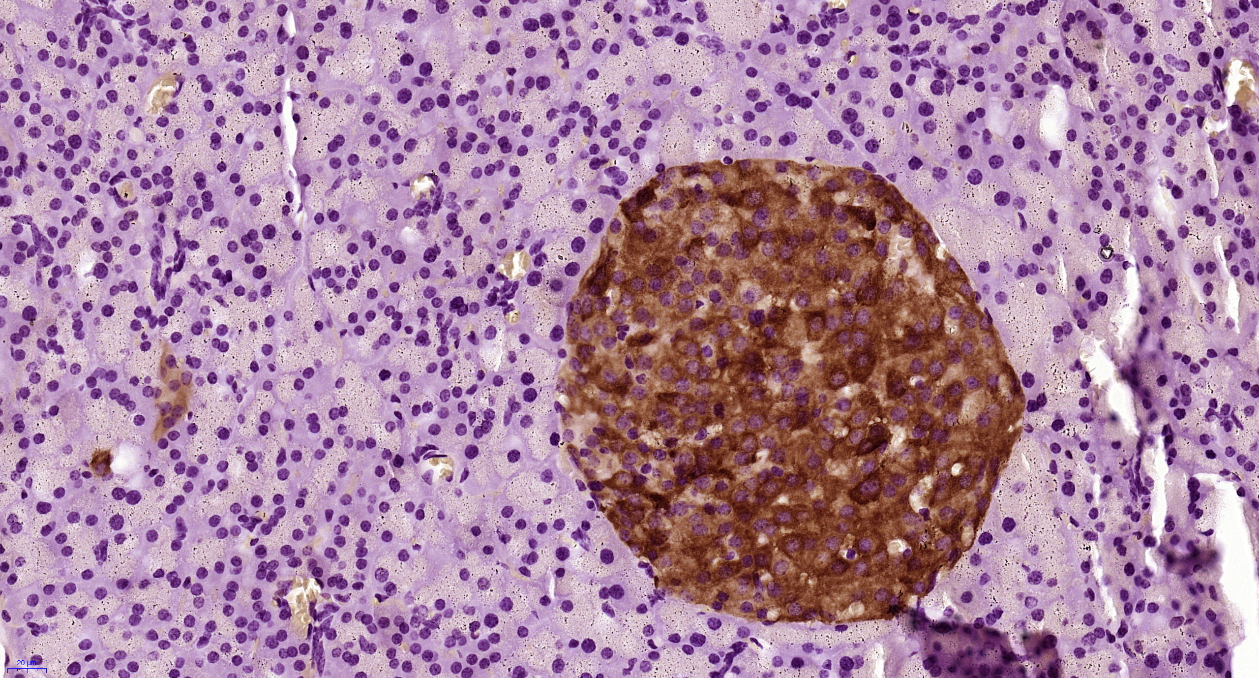
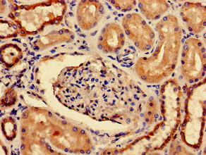
- ApplicationsImmunoHistoChemistry, ImmunoHistoChemistry Paraffin
- CAS Number26628-22-8
- CertificationResearch Use Only
- ClonalityMonoclonal
- Clone IDPRLR/3785R
- Concentration0.1 mg/ml
- ConjugateOther Conjugate
- Gene ID5618
- Target namePRLR
- Target descriptionprolactin receptor
- Target synonymsHPRL, MFAB, RI-PRLR, hPRLrI, prolactin receptor, hPRL receptor, secreted prolactin binding protein
- HostRabbit
- IsotypeIgG
- Protein IDP16471
- Protein NameProlactin receptor
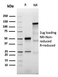
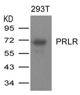
- Scientific DescriptionRecognizes a protein of 70 kDa, identified as prolactin receptor. Prolactin is a pituitary hormone involved in the stimulation of milk production, salt and water regulation, growth, development and reproduction. The initial step in its action is the binding to a specific membrane receptor (prolactin receptor), which belongs to the superfamily of class 1 cytokine receptors. The function of the prolactin receptor is mediated, at least in part, by two families of signaling molecules: Janus kinases and signal transducers and activators of transcription. Primary antibodies are available purified, or with a selection of fluorescent CF® Dyes and other labels. CF® Dyes offer exceptional brightness and photostability. Note: Conjugates of blue fluorescent dyes like CF®405S and CF®405M are not recommended for detecting low abundance targets, because blue dyes have lower fluorescence and can give higher non-specific background than other dye colors.
- SourceAnimal
- ReactivityHuman
- Storage Instruction2°C to 8°C,RT
- UNSPSC41116161
MSDS
Related products
Product group Antibodies
Anti-Prolactin Receptor Antibody188-10160
ApplicationsFlow Cytometry
ReactivityHuman
TargetPRLR
- SizePrice
Product group Antibodies

ApplicationsELISA
ReactivityHuman
TargetPRLR
- SizePrice
Product group Antibodies
References
PRLR Polyclonal AntibodyBS-6445R
ApplicationsImmunoFluorescence, Western Blot, ELISA, ImmunoCytoChemistry, ImmunoHistoChemistry, ImmunoHistoChemistry Frozen, ImmunoHistoChemistry Paraffin
ReactivityHuman, Mouse, Rat
TargetPRLR
- SizePrice
Product group Antibodies
ApplicationsImmunoPrecipitation, Western Blot, ImmunoCytoChemistry, ImmunoHistoChemistry
ReactivityMouse, Porcine, Rat
TargetPRLR
- SizePrice
Product group Antibodies
PRLR AntibodyCSB-PA018727LA01HU
ApplicationsELISA, ImmunoHistoChemistry
ReactivityHuman
TargetPRLR
- SizePrice
Product group Antibodies
Prolactin Receptor antibodyGTX50723
ApplicationsWestern Blot
ReactivityHuman
TargetPRLR
- SizePrice

Product group Antibodies
Anti-Prolactin Receptor/PRLR Antibody Picoband(r)PB9782-CARRIER-FREE
ApplicationsWestern Blot
ReactivityHuman
TargetPRLR
- SizePrice