

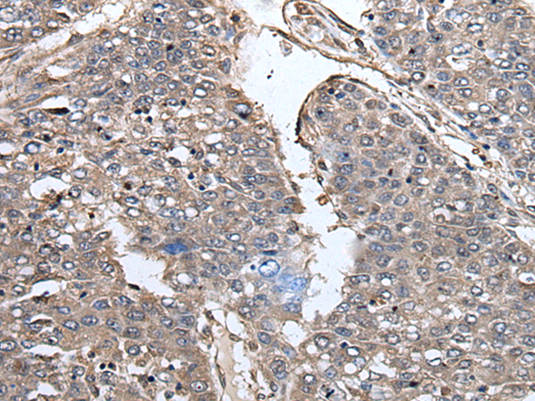
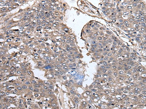
Immunohistochemistry of paraffin-embedded Human liver cancer tissue using TA364941 (PRUNE1 Antibody) at dilution 1/40 (Original magnification: x200)
Immunohistochemistry of paraffin-embedded Human liver cancer tissue using TA364941 (PRUNE1 Antibody) at dilution 1/40 (Original magnification: x200)
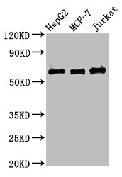
PRUNE (PRUNE1) Rabbit Polyclonal Antibody
TA364941
Overview
- SupplierOriGene
- Product NamePRUNE (PRUNE1) Rabbit Polyclonal Antibody
- Delivery Days Customer14
- ApplicationsImmunoHistoChemistry
- CertificationResearch Use Only
- ClonalityPolyclonal
- Gene ID58497
- Target namePRUNE1
- Target descriptionprune exopolyphosphatase 1
- Target synonymsDRES17; DRES-17; Drosophila-related expressed sequence 17; exopolyphosphatase PRUNE1; H-PRUNE; HTCD37; NMIHBA; protein prune homolog 1; PRUNE
- HostRabbit
- IsotypeIgG
- Protein IDQ86TP1
- Protein NameExopolyphosphatase PRUNE1
- Scientific DescriptionPRUNE1 rabbit polyclonal antibody
- Storage Instruction-20°C
- UNSPSC12352203
Related products
Product group Antibodies
Prune1 Polyclonal AntibodyCAC10478
ApplicationsImmunoFluorescence, ImmunoPrecipitation, Western Blot, ELISA, ImmunoHistoChemistry
TargetPRUNE1
- SizePrice
Product group Antibodies
PRUNE1 AntibodyCSB-PA771430LA01HU
ApplicationsImmunoFluorescence, ImmunoPrecipitation, Western Blot, ELISA, ImmunoHistoChemistry
ReactivityHuman
TargetPRUNE1
- SizePrice

Product group Antibodies
DRES17 Polyclonal AntibodyBS-6299R
ApplicationsImmunoFluorescence, Western Blot, ELISA, ImmunoCytoChemistry, ImmunoHistoChemistry, ImmunoHistoChemistry Frozen, ImmunoHistoChemistry Paraffin
TargetPRUNE1
- SizePrice
Product group Antibodies

PRUNE AntibodyLS-C831772
ApplicationsELISA, ImmunoHistoChemistry
TargetPRUNE1
- SizePrice

Product group Antibodies
Anti-PRUNE1 AntibodyHPA028411
ApplicationsWestern Blot, ImmunoCytoChemistry, ImmunoHistoChemistry
ReactivityHuman
TargetPRUNE1
- SizePrice

Product group Antibodies
Anti-PRUNE1 Antibody Picoband(r)A31798-CARRIER-FREE
ApplicationsFlow Cytometry, ImmunoFluorescence, Western Blot, ELISA, ImmunoCytoChemistry
TargetPRUNE1
- SizePrice