


ELISA analysis of antigen using GTX80396 PSIP1 antibody [6E4].
Red : Control antigen 100ng
Purple : Antigen 10ng
Green : Antigen 50ng
Blue : Antigen 100ng
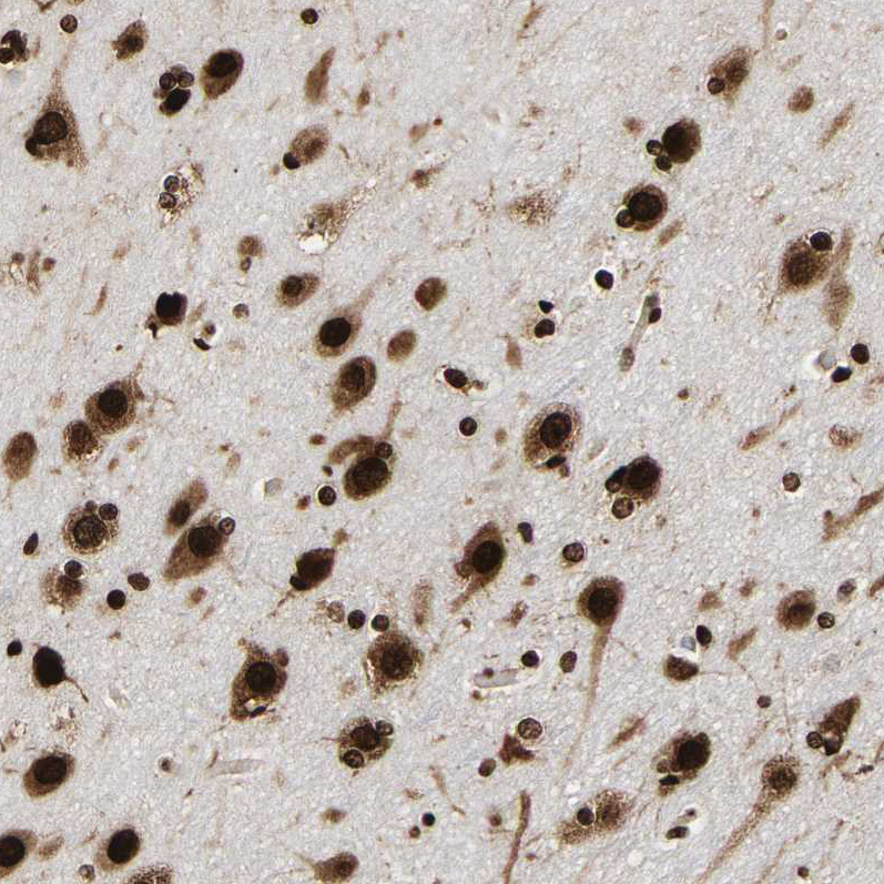
![IHC-P analysis of lung cancer tissue (left) and brain tissue (right) using GTX80396 PSIP1 antibody [6E4].](https://www.genetex.com/upload/website/prouct_img/normal/GTX80396/GTX80396_20170912_IHC-P_1_w_23061322_584.webp "IHC-P analysis of lung cancer tissue (left) and brain tissue (right) using GTX80396 PSIP1 antibody [6E4].")
![WB analysis of HepG2 (1), Jurkat (2), K562 (3), Cos7 (4), PC-12 (5), HeLa (6), and NIH3T3 (7) cell lysate using GTX80396 PSIP1 antibody [6E4].](https://www.genetex.com/upload/website/prouct_img/normal/GTX80396/GTX80396_20170912_WB_w_23061322_665.webp "WB analysis of HepG2 (1), Jurkat (2), K562 (3), Cos7 (4), PC-12 (5), HeLa (6), and NIH3T3 (7) cell lysate using GTX80396 PSIP1 antibody [6E4].")
![IHC-P analysis of breast cancer tissue (left) and ovarian cancer tissue (right) using GTX80396 PSIP1 antibody [6E4].](https://www.genetex.com/upload/website/prouct_img/normal/GTX80396/GTX80396_20170912_IHC-P_w_23061322_210.webp "IHC-P analysis of breast cancer tissue (left) and ovarian cancer tissue (right) using GTX80396 PSIP1 antibody [6E4].")
![ICC/IF analysis of NIH3T3 cells using GTX80396 PSIP1 antibody [6E4]. Green : PSIP1 Red: Actin filaments](https://www.genetex.com/upload/website/prouct_img/normal/GTX80396/GTX80396_20170912_ICCIF_w_23061322_107.webp "ICC/IF analysis of NIH3T3 cells using GTX80396 PSIP1 antibody [6E4]. Green : PSIP1 Red: Actin filaments")
ELISA analysis of antigen using GTX80396 PSIP1 antibody [6E4].
Red : Control antigen 100ng
Purple : Antigen 10ng
Green : Antigen 50ng
Blue : Antigen 100ng
PSIP1 antibody [6E4]
GTX80396
ApplicationsImmunoFluorescence, Western Blot, ELISA, ImmunoCytoChemistry, ImmunoHistoChemistry, ImmunoHistoChemistry Paraffin
Product group Antibodies
ReactivityHuman, Monkey, Mouse, Rat
TargetPSIP1
Overview
- SupplierGeneTex
- Product NamePSIP1 antibody [6E4]
- Delivery Days Customer9
- Application Supplier NoteWB: 1/500 - 1/2000. ICC/IF: 1/200 - 1/1000. IHC-P: 1/200 - 1/1000. ELISA: 1/10000. *Optimal dilutions/concentrations should be determined by the researcher.Not tested in other applications.
- ApplicationsImmunoFluorescence, Western Blot, ELISA, ImmunoCytoChemistry, ImmunoHistoChemistry, ImmunoHistoChemistry Paraffin
- CertificationResearch Use Only
- ClonalityMonoclonal
- ConjugateUnconjugated
- Gene ID11168
- Target namePSIP1
- Target descriptionPC4 and SRSF1 interacting protein 1
- Target synonymsDFS70, LEDGF, PAIP, PSIP2, p52, p75, PC4 and SFRS1-interacting protein, CLL-associated antigen KW-7, PC4 and SFRS1 interacting protein 1, dense fine speckles 70 kDa protein, lens epithelium-derived growth factor, transcriptional coactivator p52/p75
- HostMouse
- IsotypeIgG1
- Protein IDO75475
- Protein NamePC4 and SFRS1-interacting protein
- ReactivityHuman, Monkey, Mouse, Rat
- Storage Instruction-20°C or -80°C,2°C to 8°C
- UNSPSC41116161
Datasheet
Related products
Product group Antibodies

Anti-PSIP1 [RAB-C375]Ab01849-1.1
ApplicationsImmunoFluorescence, ImmunoPrecipitation
ReactivityHuman
TargetPSIP1
- SizePrice

Product group Antibodies
Anti-PSIP1 Antibody Picoband(r)A01960-2-CARRIER-FREE
ApplicationsFlow Cytometry, ImmunoFluorescence, Western Blot, ELISA, ImmunoCytoChemistry, ImmunoHistoChemistry
ReactivityHuman, Mouse, Rat
TargetPSIP1
- SizePrice
Product group Antibodies
ApplicationsFlow Cytometry, Western Blot, ImmunoCytoChemistry
ReactivityHuman, Mouse, Rat
TargetPSIP1
- SizePrice
Product group Antibodies
PSIP1 AntibodyCSB-PA018860ZA01HU
ApplicationsWestern Blot, ELISA
ReactivityHuman
TargetPSIP1
- SizePrice
Product group Antibodies
Anti-PSIP1 AntibodyHPA019697
ApplicationsChIP Chromatin ImmunoPrecipitation, ImmunoCytoChemistry, ImmunoHistoChemistry
ReactivityHuman
TargetPSIP1
- SizePrice
Product group Antibodies

PSIP1 / LEDGF AntibodyLS-C749833
ApplicationsWestern Blot, ImmunoHistoChemistry
ReactivityHuman, Mouse, Rat
TargetPSIP1
- SizePrice