

PSMD10 antibody
GTX32820
ApplicationsImmunoFluorescence, Western Blot, ImmunoCytoChemistry, ImmunoHistoChemistry, ImmunoHistoChemistry Paraffin
Product group Antibodies
ReactivityHuman, Mouse, Rat
TargetPSMD10
Overview
- SupplierGeneTex
- Product NamePSMD10 antibody
- Delivery Days Customer9
- Application Supplier NoteWB: 1:500 - 1:2000. ICC/IF: 1:50 - 1:200. IHC-P: 1:50 - 1:200. *Optimal dilutions/concentrations should be determined by the researcher.Not tested in other applications.
- ApplicationsImmunoFluorescence, Western Blot, ImmunoCytoChemistry, ImmunoHistoChemistry, ImmunoHistoChemistry Paraffin
- CertificationResearch Use Only
- ClonalityPolyclonal
- ConjugateUnconjugated
- Gene ID5716
- Target namePSMD10
- Target descriptionproteasome 26S subunit, non-ATPase 10
- Target synonymsdJ889N15.2, p28, p28(GANK), 26S proteasome non-ATPase regulatory subunit 10, 26S proteasome regulatory subunit p28, ankyrin repeat protein, gankyrin, hepatocellular carcinoma-associated protein p28-II, proteasome (prosome, macropain) 26S subunit, non-ATPase, 10
- HostRabbit
- IsotypeIgG
- Protein IDO75832
- Protein Name26S proteasome non-ATPase regulatory subunit 10
- Scientific DescriptionThis gene encodes a subunit of the PA700/19S complex, which is the regulatory component of the 26S proteasome. The 26S proteosome complex is required for ubiquitin-dependent protein degradation. This protein is a non-ATPase subunit that may be involved in protein-protein interactions. Aberrant expression of this gene may paly a role in tumorigenesis. Two transcripts encoding different isoforms have been described. Pseudogenes have been identified on chromosomes 3 and 20.[provided by RefSeq, Mar 2011]
- ReactivityHuman, Mouse, Rat
- Storage Instruction-20°C or -80°C,2°C to 8°C
- UNSPSC12352203
Datasheet
Related products
Product group Antibodies
Anti-PSMD10 (C-term) Antibody102-21934
ApplicationsWestern Blot
TargetPSMD10
- SizePrice
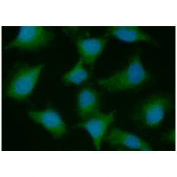
![PSMD10 antibody [N1C3] detects PSMD10 protein at cytoplasm and nucleus by immunofluorescent analysis. Sample: HepG2 cells were fixed in 4% paraformaldehyde at RT for 15 min. Green: PSMD10 protein stained by PSMD10 antibody [N1C3] (GTX113813) diluted at 1:500. Blue: Hoechst 33342 staining. Scale bar = 10 μm.](https://www.genetex.com/upload/website/prouct_img/normal/GTX113813/GTX113813_40150_20141212_IFA_w_23060501_718.webp)
Product group Antibodies
PSMD10 antibody [N1C3]GTX113813
ApplicationsImmunoFluorescence, Western Blot, ImmunoCytoChemistry
ReactivityHuman
TargetPSMD10
- SizePrice
Product group Antibodies
PSMD10 antibody [AT1F8]GTX57642
ApplicationsImmunoFluorescence, Western Blot, ImmunoCytoChemistry
ReactivityHuman
TargetPSMD10
- SizePrice
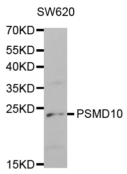
![WB analysis of various cell lines using GTX83773 PSMD10 antibody [3F6]. Loading : 35 ug per lane](https://www.genetex.com/upload/website/prouct_img/normal/GTX83773/GTX83773_3899_WB_w_23061420_327.webp)
Product group Antibodies
PSMD10 antibody [3F6]GTX83773
ApplicationsFlow Cytometry, Western Blot
ReactivityHuman
TargetPSMD10
- SizePrice
Product group Antibodies
ApplicationsWestern Blot, ELISA, ImmunoCytoChemistry, ImmunoHistoChemistry, ImmunoHistoChemistry Frozen, ImmunoHistoChemistry Paraffin
TargetPSMD10
- SizePrice
Product group Antibodies
PSMD10 AntibodyCSB-PA018899LA01HU
ApplicationsImmunoFluorescence, ELISA, ImmunoHistoChemistry
ReactivityHuman
TargetPSMD10
- SizePrice
Product group Antibodies

PSMD10 / Gankyrin AntibodyLS-C769924
ApplicationsELISA, ImmunoHistoChemistry
ReactivityHuman, Mouse, Rat
TargetPSMD10
- SizePrice
Product group Antibodies
Anti-PSMD10 AntibodyA30531
ApplicationsImmunoFluorescence, Western Blot, ImmunoHistoChemistry
ReactivityHuman, Mouse, Rat
- SizePrice