

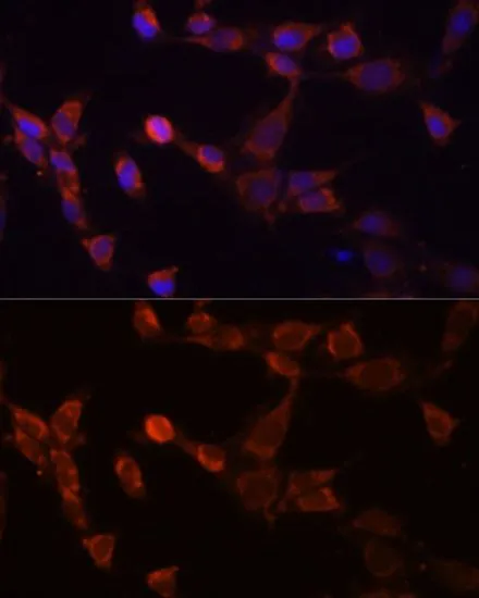
ICC/IF analysis of NIH/3T3 cells using GTX64996 PSMD14 antibody. Blue : DAPI Dilution : 1:100

ICC/IF analysis of NIH/3T3 cells using GTX64996 PSMD14 antibody. Blue : DAPI Dilution : 1:100
PSMD14 antibody
GTX64996
ApplicationsImmunoFluorescence, Western Blot, ImmunoCytoChemistry
Product group Antibodies
ReactivityHuman, Mouse
TargetPSMD14
Overview
- SupplierGeneTex
- Product NamePSMD14 antibody
- Delivery Days Customer9
- Application Supplier NoteWB: 1:500 - 1:2000. ICC/IF: 1:50 - 1:100. *Optimal dilutions/concentrations should be determined by the researcher.Not tested in other applications.
- ApplicationsImmunoFluorescence, Western Blot, ImmunoCytoChemistry
- CertificationResearch Use Only
- ClonalityPolyclonal
- ConjugateUnconjugated
- Gene ID10213
- Target namePSMD14
- Target descriptionproteasome 26S subunit, non-ATPase 14
- Target synonymsPAD1, POH1, RPN11, 26S proteasome non-ATPase regulatory subunit 14, 26S proteasome regulatory subunit rpn11, 26S proteasome-associated PAD1 homolog 1, proteasome (prosome, macropain) 26S subunit, non-ATPase, 14, testis tissue sperm-binding protein Li 69n
- HostRabbit
- IsotypeIgG
- Protein IDO00487
- Protein Name26S proteasome non-ATPase regulatory subunit 14
- Scientific DescriptionThis gene encodes a component of the 26S proteasome. The 26S proteasome is a large multiprotein complex that catalyzes the degradation of ubiquitinated intracellular proteins. The encoded protein is a component of the 19S regulatory cap complex of the 26S proteasome and mediates substrate deubiquitination. A pseudogene of this gene is also located on the long arm of chromosome 2. [provided by RefSeq, Feb 2012]
- ReactivityHuman, Mouse
- Storage Instruction-20°C or -80°C,2°C to 8°C
- UNSPSC41116161
Datasheet
Related products
Product group Antibodies
PSMD14 AntibodyCSB-PA018904OA01HU
ApplicationsImmunoFluorescence, Western Blot, ELISA, ImmunoHistoChemistry
ReactivityHuman, Rat
TargetPSMD14
- SizePrice
Product group Antibodies
Anti-PSMD14 AntibodyA11351
ApplicationsImmunoFluorescence, Western Blot, ImmunoCytoChemistry
ReactivityHuman, Mouse, Rat
- SizePrice

Product group Antibodies
Anti-POH1/PSMD14 Antibody Picoband(r)A06584-1-CARRIER-FREE
ApplicationsFlow Cytometry, ImmunoFluorescence, Western Blot, ELISA, ImmunoCytoChemistry, ImmunoHistoChemistry
ReactivityHuman, Mouse, Rat
TargetPSMD14
- SizePrice

Product group Antibodies
Anti-PSMD14 AntibodyHPA002114
ApplicationsWestern Blot, ImmunoCytoChemistry, ImmunoHistoChemistry
ReactivityHuman, Mouse, Rat
TargetPSMD14
- SizePrice
Product group Antibodies

PSMD14 AntibodyLS-C497489
ApplicationsWestern Blot
ReactivityHuman, Mouse
TargetPSMD14
- SizePrice
Product group Antibodies
PSMD14 Polyclonal AntibodyCAC15721
ApplicationsImmunoFluorescence, Western Blot, ELISA, ImmunoHistoChemistry
ReactivityRat
TargetPSMD14
- SizePrice
Product group Antibodies
PSMD14 Recombinant AntibodyBSM-62064R
ApplicationsImmunoFluorescence, Western Blot, ImmunoCytoChemistry, ImmunoHistoChemistry, ImmunoHistoChemistry Frozen, ImmunoHistoChemistry Paraffin
ReactivityHuman, Mouse, Rat
TargetPSMD14
- SizePrice
Product group Antibodies
Anti-PSMD14 Antibody144-10782
ApplicationsWestern Blot
ReactivityHuman, Mouse
TargetPSMD14
- SizePrice