

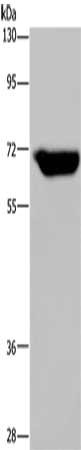
PSR antibody
GTX10526
ApplicationsImmunoPrecipitation, Western Blot
Product group Antibodies
ReactivityHuman, Mouse
TargetJMJD6
Overview
- SupplierGeneTex
- Product NamePSR antibody
- Delivery Days Customer9
- Application Supplier NoteWB: 1:500~1:1000. *Optimal dilutions/concentrations should be determined by the researcher.Not tested in other applications.
- ApplicationsImmunoPrecipitation, Western Blot
- CertificationResearch Use Only
- ClonalityPolyclonal
- Concentration1-1.5 mg/ml
- ConjugateUnconjugated
- Gene ID23210
- Target nameJMJD6
- Target descriptionjumonji domain containing 6, arginine demethylase and lysine hydroxylase
- Target synonymsPSR, PTDSR, PTDSR1, bifunctional arginine demethylase and lysyl-hydroxylase JMJD6, arginine demethylase and lysine hydroxylase, histone arginine demethylase JMJD6, jmjC domain-containing protein 6, jumonji domain-containing protein 6, lysyl-hydroxylase JMJD6, peptide-lysine 5-dioxygenase JMJD6, phosphatidylserine receptor
- HostRabbit
- IsotypeIgG
- Protein IDQ6NYC1
- Protein NameBifunctional arginine demethylase and lysyl-hydroxylase JMJD6
- Scientific DescriptionMost cells that undergo apoptosis are rapidly recognized, engulfed, and digested by professional phagocytes or by neighboring cells and eliminated from the organism. In this way, tissue exposure to potentially cytotoxic, immunogenic, or inflammatory cellular contents is prevented. As a result neither inflammation nor inappropriate immune responses are induced in this process. Removal of apoptotic cells comprise the following steps: surface alteration and ligand expression on the apoptotic cell, ligand recognition by tethering receptors on the phagocyte, initiation of appropriate signaling pathways in the phagocyte, engulfment and finally, digestion. Various molecules including proteins, carbohydrates, and phospholipids have been suggested to act as cell surface exposed markers for recognition by phagocytes. Virtually all apoptotic cells permanently express phosphatidylserine (PS) on the outer leaflet of their plasma membrane. Such loss of phospholipids asymmetry and exposure of PS does not occur in resting cells and appears only transiently following activation of certain cells. Phosphatidylserine Receptor (PSR) has been reported and shown to recognize phosphatidylserine in a stereospecific manner. PSR is an evolutionarily conserved glycoprotein that is widely expressed in cells and tissues. It is very susceptible to cleavage by protease or elastase. PSR engagement plays an important role in macropinocytosis and uptake of tethered apoptotic cells. Cooperation with other receptors and their ligands may also be needed for ingestion of apoptotic cells. PSR is also involved in suppression of inflammatory and immune responses.
- ReactivityHuman, Mouse
- Storage Instruction-20°C or -80°C,2°C to 8°C
- UNSPSC12352203
Datasheet
Related products
Product group Antibodies
JMJD6 AntibodyCSB-PA163798
ApplicationsWestern Blot, ELISA
ReactivityHuman, Mouse, Rat
TargetJMJD6
- SizePrice
Product group Antibodies
ApplicationsImmunoPrecipitation, Western Blot, ImmunoCytoChemistry, ImmunoHistoChemistry
ReactivityMouse, Rat
TargetJMJD6
- SizePrice
Product group Antibodies

Anti-JMJD6 [APSR-14.4]Ab00928-10.0
ApplicationsImmunoFluorescence, ImmunoPrecipitation, Western Blot, Other Application
ReactivityHuman
TargetJMJD6
- SizePrice
Product group Antibodies
Anti-JMJD6 AntibodyA31666
ApplicationsImmunoFluorescence, ImmunoPrecipitation, Western Blot, ChIP Chromatin ImmunoPrecipitation, ImmunoHistoChemistry
ReactivityHuman, Mouse, Rat
- SizePrice
Product group Antibodies
Anti-JMJD6 Antibody144-66010
ApplicationsImmunoFluorescence, Western Blot, ImmunoHistoChemistry
ReactivityHuman, Mouse, Rat
TargetJMJD6
- SizePrice

Product group Antibodies
Anti-JMJD6 AntibodyHPA059156
ApplicationsImmunoCytoChemistry
ReactivityHuman
TargetJMJD6
- SizePrice
Product group Antibodies

JMJD6 / PSR AntibodyLS-C401786
ApplicationsWestern Blot, ELISA
ReactivityHuman, Mouse, Rat
TargetJMJD6
- SizePrice
Product group Antibodies
JMJD6 Polyclonal AntibodyBS-6842R
ApplicationsImmunoFluorescence, Western Blot, ELISA, ImmunoCytoChemistry, ImmunoHistoChemistry, ImmunoHistoChemistry Frozen, ImmunoHistoChemistry Paraffin
ReactivityBovine, Canine, Chicken, Equine, Human, Mouse, Porcine, Rabbit, Rat, Sheep, Zebra Fish
TargetJMJD6
- SizePrice
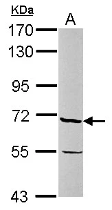
Product group Antibodies
PSR antibodyGTX110746
ApplicationsWestern Blot, ImmunoHistoChemistry, ImmunoHistoChemistry Paraffin
ReactivityHuman, Mouse
TargetJMJD6
- SizePrice