

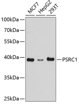
WB analysis of various sample lysates using GTX32823 PSRC1 antibody. Dilution : 1:1000 Loading : 25μg per lane


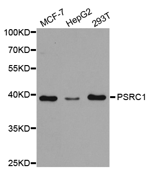
WB analysis of various sample lysates using GTX32823 PSRC1 antibody. Dilution : 1:1000 Loading : 25μg per lane
PSRC1 antibody
GTX32823
ApplicationsWestern Blot, ImmunoHistoChemistry, ImmunoHistoChemistry Paraffin
Product group Antibodies
ReactivityHuman, Mouse
TargetPSRC1
Overview
- SupplierGeneTex
- Product NamePSRC1 antibody
- Delivery Days Customer9
- Application Supplier NoteWB: 1:500 - 1:2000. IHC-P: 1:50 - 1:100. *Optimal dilutions/concentrations should be determined by the researcher.Not tested in other applications.
- ApplicationsWestern Blot, ImmunoHistoChemistry, ImmunoHistoChemistry Paraffin
- CertificationResearch Use Only
- ClonalityPolyclonal
- ConjugateUnconjugated
- Gene ID84722
- Target namePSRC1
- Target descriptionproline and serine rich coiled-coil 1
- Target synonymsDDA3, FP3214, proline/serine-rich coiled-coil protein 1, differential display and activated by p53, p53-regulated DDA3, proline/serine-rich coiled-coil 1
- HostRabbit
- IsotypeIgG
- Protein IDQ6PGN9
- Protein NameProline/serine-rich coiled-coil protein 1
- Scientific DescriptionThis gene encodes a proline-rich protein that is a target for regulation by the tumor suppressor protein p53. The encoded protein plays an important role in mitosis by recruiting and regulating microtubule depolymerases that destabalize microtubules. Alternatively spliced transcript variants encoding different isoforms have been described. [provided by RefSeq, Apr 2014]
- ReactivityHuman, Mouse
- Storage Instruction-20°C or -80°C,2°C to 8°C
- UNSPSC41116161
Datasheet
Related products
Product group Antibodies
Anti-PSRC1 AntibodyA30797
ApplicationsWestern Blot, ImmunoHistoChemistry
ReactivityHuman
- SizePrice
Product group Antibodies
Anti-PSRC1 AntibodyA03961
ApplicationsImmunoFluorescence, Western Blot, ImmunoCytoChemistry
ReactivityHuman, Mouse, Rat
TargetPSRC1
- SizePrice
Product group Antibodies
Anti-PSRC1 Antibody144-05484
ApplicationsWestern Blot, ImmunoHistoChemistry
ReactivityHuman, Mouse
TargetPSRC1
- SizePrice
Product group Antibodies
References
PSRC1 Polyclonal AntibodyBS-7619R
ApplicationsImmunoFluorescence, Western Blot, ELISA, ImmunoCytoChemistry, ImmunoHistoChemistry, ImmunoHistoChemistry Frozen, ImmunoHistoChemistry Paraffin
ReactivityBovine, Canine, Human, Mouse, Porcine, Rabbit, Rat
TargetPSRC1
- SizePrice
Product group Antibodies

PSRC1 / DDA3 AntibodyLS-C334083
ApplicationsWestern Blot, ImmunoHistoChemistry
ReactivityHuman, Mouse
TargetPSRC1
- SizePrice

Product group Antibodies
Anti-PSRC1 AntibodyHPA056561
ApplicationsImmunoCytoChemistry
ReactivityHuman
TargetPSRC1
- SizePrice
![PSRC1 antibody detects PSRC1 protein by immunohistochemical analysis. Sample: Frozen-sectioned mouse mouse cerebellum. Green: PSRC1 stained by PSRC1 antibody (GTX128047) diluted at 1:250. Red: NF-H, stained by NF-H antibody [GT114] (GTX634289) diluted at 1:500. Blue: Fluoroshield with DAPI (GTX30920).](https://www.genetex.com/upload/website/prouct_img/normal/GTX128047/GTX128047_41213_20180530_IHC-Fr_M_2_w_23060522_851.webp)
Product group Antibodies
PSRC1 antibodyGTX128047
ApplicationsImmunoFluorescence, Western Blot, ImmunoCytoChemistry, ImmunoHistoChemistry, ImmunoHistoChemistry Frozen, ImmunoHistoChemistry Paraffin
ReactivityHuman, Mouse
TargetPSRC1
- SizePrice
![PSRC1 antibody [GT615] detects PSRC1 protein at cytoplasm by immunofluorescent analysis. Sample: Mock and transfected 293T cells were fixed in 4% paraformaldehyde at RT for 15 min. Green: PSRC1 stained by PSRC1 antibody [GT615] (GTX628833) diluted at 1:2000. Blue: Hoechst 33342 staining. Scale bar= 10 μm.](https://www.genetex.com/upload/website/prouct_img/normal/GTX628833/GTX628833_41211_20180801_ICC_IF_B_w_23061202_158.webp)
Product group Antibodies
PSRC1 antibody [GT615]GTX628833
ApplicationsImmunoFluorescence, Western Blot, ImmunoCytoChemistry
ReactivityHuman
TargetPSRC1
- SizePrice
![PSRC1 antibody [GT982] detects PSRC1 protein by Western blot analysis. A. 30 μg 293T whole cell lysate/extract B. 30 μg whole cell lysate/extract of human PSRC1-transfected 293T cells 10 % SDS-PAGE PSRC1 antibody [GT982] (GTX629077) dilution: 1:5000](https://www.genetex.com/upload/website/prouct_img/normal/GTX629077/GTX629077_41295_WB_B_w_23051500_158.webp)
Product group Antibodies
PSRC1 antibody [GT982]GTX629077
ApplicationsImmunoFluorescence, Western Blot, ImmunoCytoChemistry
ReactivityHuman
TargetPSRC1
- SizePrice
Product group Antibodies
Anti-PSRC1 AntibodyCAB5484
ApplicationsWestern Blot, ELISA
ReactivityHuman
TargetPSRC1
- SizePrice